41 neuron labels
Neuron Label Flashcards | Quizlet Start studying Neuron Label. Learn vocabulary, terms, and more with flashcards, games, and other study tools. › articles › s41586/022/04711-3Transcriptomic mapping uncovers Purkinje neuron plasticity ... May 11, 2022 · a, AAV-Plcb4-tdTomato labels Plcb4+ Purkinje neurons transduced by Purkinje neuron-specific GCaMP (AAV-L7-cre + rAAV-CAG-FLEx-jGCaMP7f). Scale bar, 50 μm. Scale bar, 50 μm. Similar results were ...
Nerve Cell (Neuron) Labeling Page - Exploring Nature The style of citing shown here is from the MLA Style Citations (Modern Language Association). When citing a WEBSITE the general format is as follows. Author Last Name, First Name (s). "Title: Subtitle of Part of Web Page, if appropriate." Title: Subtitle: Section of Page if appropriate. Sponsoring/Publishing Agency, If Given.
Neuron labels
Neuron Label Worksheets - K12 Workbook Neuron Label Displaying all worksheets related to - Neuron Label . Worksheets are Neuron anatomy activity, Work for classes 1 and 2, Neurotransmission fact, Neuron labeling work answers, Being brainy activity pack, Levels of organization foldable, Chapter 12 central nervous system, Brain anatomy. › pmc › articlesFunctional network organization of the human brain - PMC Nov 17, 2011 · Neuron. 2010a; 67:156–170. [PMC free article] [Google Scholar] Nelson SM, Dosenbach NU, Cohen AL, Wheeler ME, Schlaggar BL, Petersen SE. Role of the anterior insula in task-level control and focal attention. Brain Struct Funct. 2010b; 214:669–680. [PMC free article] [Google Scholar] Newman MEJ. Networks: An introduction. sfponline.org › Uploads › 71NERVOUS SYSTEM WORKSHEET - St. Francis Preparatory School 1. The diagram below is of a nerve cell or neuron. Add the following labels to the diagram: Axon Myelin sheath Cell body Dendrites Muscle fibers Axon terminals 2. Color in the diagram as suggested below. Axon - purple Axon Terminals – orange Myelin sheath – yellow Cell body – blue Dendrites – green Muscle fibers - red 3.
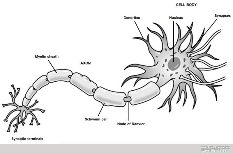
Neuron labels. Labels · vnasyj/ineuron · GitHub Contribute to vnasyj/ineuron development by creating an account on GitHub. Neuron (Nerve Cell) Types, Structure and Function Neurons, also known as nerve cells, are essentially the cells that make up the brain and the nervous system. Neurons do not touch each other, but where one neuron comes close to another neuron, a synapse is formed between the two. The function of a neuron is to transmit nerve impulses along the length of an individual neuron and across the ... Neuron Label - The Biology Corner Neuron Label. Publisher: Biologycorner.com; This work is licensed under a Creative Commons Attribution-NonCommercial 3.0 Unported License. This picture of the neuron is unlabeled, write in the labels to test your knowledge of the anatomy of a neuron. Neuron Label. ... Label Neuron Anatomy Printout - EnchantedLearning.com Read the definitions, then label the neuron diagram below. axon - the long extension of a neuron that carries nerve impulses away from the body of the cell. cell body - the cell body of the neuron; it contains the nucleus (also called the soma) dendrites - the branching structure of a neuron that receives messages (attached to the cell body)
› pmc › articlesThe Function of α-Synuclein - PMC Sep 18, 2013 · In brainstem-type Lewy bodies, the pale-staining halo, which contains filaments by electron microscopy, labels more strongly for α-synuclein than the acidophilic core (Goedert et al., 2013). Dystrophic neurites and the less discrete cortical-type Lewy bodies contain similar filaments (Marui et al., 2002). Although Lewy bodies were originally ... Neuron Diagram & Types | Ask A Biologist Nerve Cell: Dendrites receive messages from other neurons. The message then moves through the axon to the other end of the neuron, then to the tips of the axon and then into the space between neurons. From there the message can move to the next neuron. Neurons pass messages to each other using a special type of electrical signal. Neuron Labeling - The Biology Corner Neuroglia Labeling. Shannan Muskopf December 22, 2020. This labeling activity was created for remote learning during the 2020 pandemic. Students in anatomy and physiology spent this year learning remotely, which required some adjustments in how material was presented. In the past, students would label a nerve cell and color neuroglia cells ... › articles › s41593/022/01041-5Single-neuron projectome of mouse prefrontal cortex | Nature ... Mar 31, 2022 · a, The pipeline of PFC single-neuron projectome reconstruction including sparse labeling, fMOST imaging, image compression using FNT-slice2cube, neurite tracing using FNT-tracer, quality control ...
A Labelled Diagram of Neuron with Detailed decription A neuron is a type of cell that is largely responsible for conveying information via electrical and chemical impulses. The brain, spinal cord, and peripheral nerves all contain them. The nerve cell is another name for a neuron. The structure of a neuron changes depending on its form and size, as well as its function and location. Neuron Markers - BioLegend Neurons have highly compartmentalized structures that are generally classified into soma (cell body), axon, dendrite, axonal terminal, and synapse. BioLegend offers antibodies against markers that are expressed in each structural unit of a neuron, and allow their identification using applications such as microscopy, including immunohistochemistry (IHC) or immunocytochemistry (ICC). A Guide to Understand Neuron with Neuron Diagram | EdrawMax Online 3. How to Draw a Neuron Diagram To learn about the structure of the neurons, the students can use a neuron labeled diagram. The students may follow these steps to make their neuron diagram, but the process is complex: 3.1 How to Draw a Neuron Diagram from Sketch Step 1: First, the students need to draw a circle. Based on it, they need to draw a ... What Is a Neuron? Diagrams, Types, Function, and More Takeaway. Neurons, also known as nerve cells, send and receive signals from your brain. While neurons have a lot in common with other types of cells, they're structurally and functionally unique ...
The Anatomy and Physiology of Animals/Nervous System Worksheet ...
› doi › 10A million spiking-neuron integrated circuit with a scalable ... Aug 08, 2014 · A memory (static random-access memory) stores all the data for each neuron, a time-multiplexed neuron circuit updates neuron membrane potentials, a scheduler buffers incoming spike events to implement axonal delays, a router relays spike events, and an event-driven controller orchestrates the core’s operation.
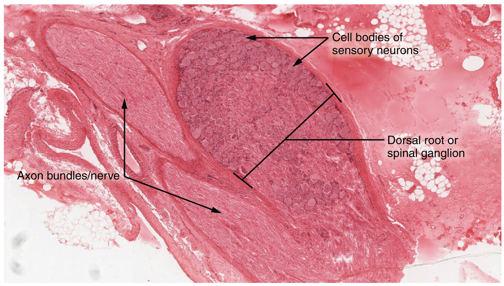
This micrograph shows the structure of the dorsal root ganglion. The ...
Neuron Label Activity - StuDocu Name each part of the neuron Explain the function of each part. Dendrites Receive messages from other cells. They look like trees, so their "branches" stem on each side of the neuron so that it can detect the information. Cell Body The cell body is the bulky part of the neuron because that's where everything is held.
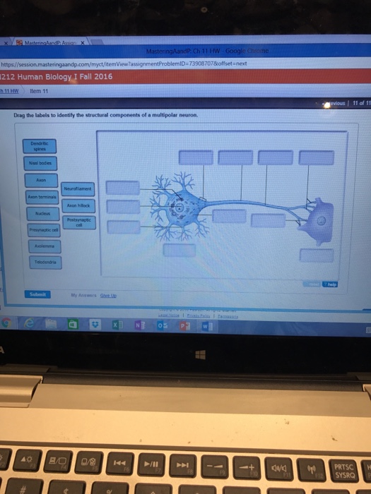
Solved: Drag The Labels To Identify The Structural Compone... | Chegg.com
Neuron Label Neuron Label. This picture of the neuron is unlabeled, write in the labels to test your knowledge of the anatomy of a neuron. Biologycorner. 17k followers. Teaching Biology. Science Biology. Life Science. Ap Biology. Computer Science. Human Body Unit. Human Body Systems. Brain Nervous System. Ap Psychology ...
Print Chapter 11 - Neurophysiology Activities flashcards | Easy Notecards
Neuron Labelling Teaching Resources | Teachers Pay Teachers This interactive Slides activity focuses on the parts/organelles in a typical nerve cell (neuron). The post actually contains three assignments all-in-one. Slide #1 is a drag-and-drop, slide #2 involves labeling with a word bank and clickable text boxes, and slide #3 challenges students label the parts of the neuron but does not provide a word ...

| In utero electroporation (IUE) selectively labels cortical neurons in ...
Pics Of Labeled Of A Neuron Pictures, Images and Stock Photos Motor neuron, detailed and accurate, labeled. The nervous system The human nervous system vector medical illustration pics of labeled of a neuron stock illustrations. The nervous system. Dendritic cells vector illustration. Anatomical labeled closeup scheme with progenitor, immature, nucleus and membrane extensions.
Brain Anatomy Image Gallery - DynamicBrain
Neurons - Neuron (labels) - histology slide Neuron (labels) - histology slide. This is a histology slide of a neuron. Histology slide courtesy of William L. Todt, Ph.D. at Concordia College, Moorhead, Minnesota. File information.
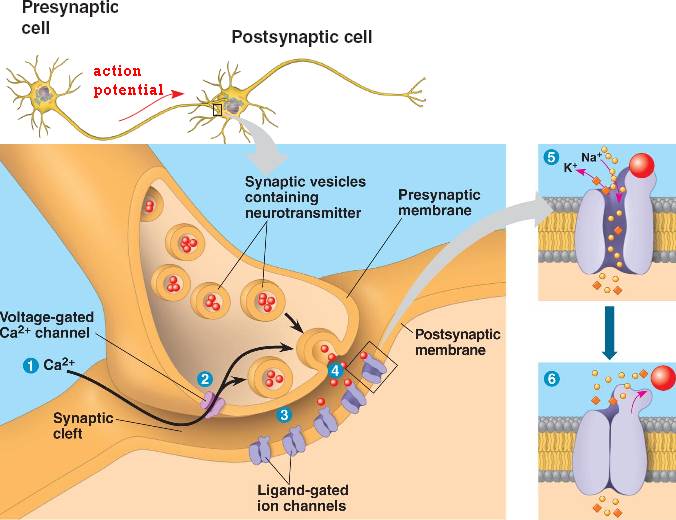
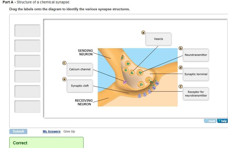
synapse.html 48_17ChemicalSynapse.jpg
Neuron - Label - Labelled diagram Neuron - Label. Share Share by Aspillane. Y12 Y13 Psychology. Like. Edit Content. Embed. More. Leaderboard. Show more Show less . This leaderboard is currently private. Click Share to make it public. This leaderboard has been disabled by the resource owner. This leaderboard is disabled as your options are different to the resource owner. ...

Label the Neuron
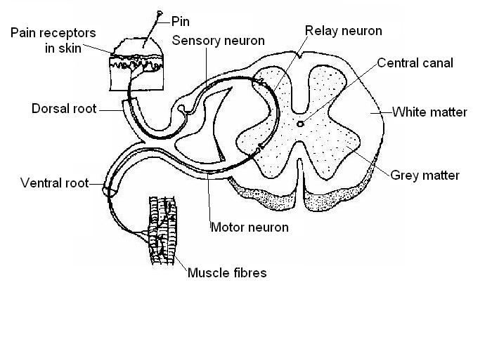
wikieducator.org › Nervous_System_Worksheet_AnswersNervous System Worksheet Answers - WikiEducator Jan 14, 2008 · B. Sensory neuron: The nerve cell that carries impulses from a sense receptor to the brain or spinal cord. C. Relay neuron: The nerve cell that connects sensory and motor neurons A. Motor neuron: The nerve cell that transmits impulses from the brain or spinal cord to a muscle or gland
Examination of hippocampal cell types using RNA-seq | Janelia Research ...
What Is a Neuron? - Definition, Structure, Parts and Function What is a Neuron? Neurons are the building blocks of the nervous system. They receive and transmit signals to different parts of the body. This is carried out in both physical and electrical forms. There are several different types of neurons that facilitate the transmission of information. The sensory neurons carry information from the sensory ...
Post a Comment for "41 neuron labels"