42 label the photomicrograph of thin skin.
Photomicrograph of Thin Skin Quiz - PurposeGames.com This is an online quiz called Photomicrograph of Thin Skin. There is a printable worksheet available for download here so you can take the quiz with pen and paper. Your Skills & Rank. Total Points. 0. Get started! Today's Rank--0. Today 's Points. One of us! Game Points. 5. Lab 9: Pre-Lab Homework Flashcards - Quizlet Label the photomicrograph of thin skin. In general, nerves from the posterior division of the brachial plexus tend to innervate muscles that extend the parts of the upper limb. True/False True Match the label to its appropriate spinal cord component. -dorsal root ganglion -white matter -gray matter -epidural space -dorsal ramus
Label the photomicrograph in Figure 7.4. Examine a slide of hairy skin ... Label The Photomicrograph Of The Skin And Its Accessory Structures. Sebaceous Gland Duct Of Sebaceous Gland Epidermis Hair Follicle ... Activity 4 Differentiating Sebaceous and Sweat Glands Microscopically Using the slide thin skin with hairs and the photomicrographs of cutaneous glands (Figure 7.6) as a guide, identify sebaceous and eccrine ...

Label the photomicrograph of thin skin.
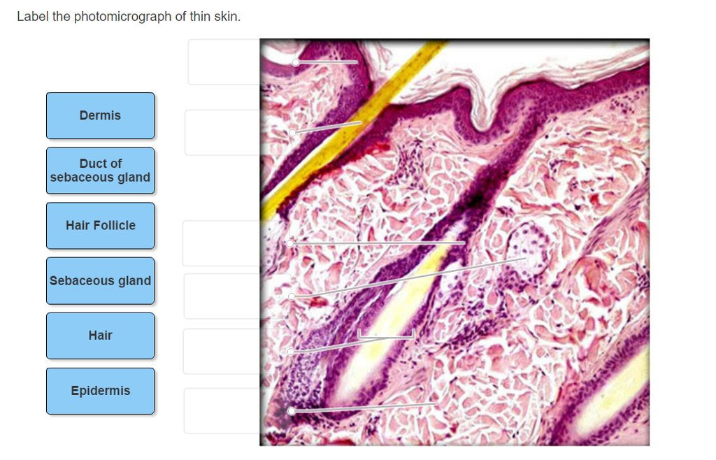
Question : Question 31 points Label the photomicrograph of thin skin ... Question : Question 31 points Label the photomicrograph of thin skin. Hair Follicle : 391984. Question. 31 points Label the photomicrograph of thin skin. Hair Follicle Hair Dermis Sebaceous gland Duct of sebaceous gland Reset zoom. Solution. 5 (1 Ratings ) Solved. Biology 2 Years Ago 68 Views. unit 4 lab.docx - LAB Unit 4 EXERCISE 7: The ... - Course Hero FIGURE 7.4: Diagram of the skin and accessory structures. • apocrine (AP-oh-krin) sweat gland • arrector pili (PIE-lee) muscle • eccrine (EK-rin) sweat gland • hair bulb • hair follicle • hair root • hair shaft • papilla (puh-PILL-uh) of hair • sebaceous (se-BAY-shus) gland 1. Hair shaft 2. Hair root 3. Sebaceous glands 4. Arrector pili muscle 5. Label The Photomicrograph Of Thin Skin Quizlet - Skin Labeling Review ... Label the photomicrograph of thin skin. Label the photomicrograph of thick skin. D) stratum corneum has fewer layers in. Start studying photomicrographs of skin (thin skin). Learn vocabulary, terms, and more with flashcards, games, and other study tools. C) contains more sweat glands than thin skin. Label the photomicrograph of thin skin.
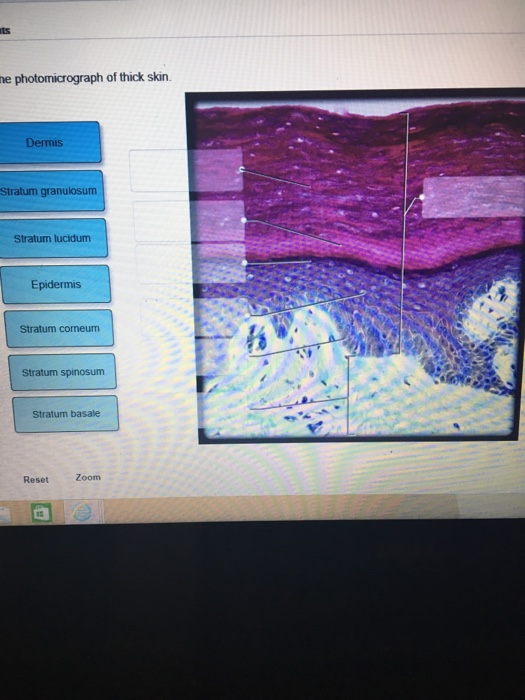
Label the photomicrograph of thin skin.. 5.1 Layers of the Skin - Anatomy & Physiology Skin that has four layers of cells is referred to as "thin skin.". From deep to superficial, these layers are the stratum basale, stratum spinosum, stratum granulosum, and stratum corneum. Most of the skin can be classified as thin skin. "Thick skin" is found only on the palms of the hands and the soles of the feet. Layers of the Skin - Anatomy and Physiology Skin that has four layers of cells is referred to as "thin skin.". From deep to superficial, these layers are the stratum basale, stratum spinosum, stratum granulosum, and stratum corneum. Most of the skin can be classified as thin skin. "Thick skin" is found only on the palms of the hands and the soles of the feet. Label The Photomicrograph Of Thin Skin. : Chapter 13, Page 3 ... Academia.edu is a platform for academics to share research papers. Label The Photomicrograph Of Thin Skin. : Chapter 13, Page 3 - HistologyOLM. 28.09.2020 · this involves depositing a thin layer of heavy metal (eg, platinum) on the specimen by placing it in the path of a beam of metal ions in a vacuum. The beam is directed at a low angle to ... Anatomy, Skin (Integument), Epidermis - StatPearls - NCBI Bookshelf Skin is the largest organ in the body and covers the body's entire external surface. It is made up of three layers, the epidermis, dermis, and the hypodermis, all three of which vary significantly in their anatomy and function. The skin's structure is made up of an intricate network which serves as the body's initial barrier against pathogens, UV light, and chemicals, and mechanical injury ...
PDF Name the Condition - Dr. Scott Croes' Website Name the 4 layers of thin skin in both the cartoon and the photomicrograph. Name the 4 layers of thin skin in both the cartoon and the photomicrograph. •Name the Layers of skin and label the dermal papilla and dermis •Name the Layers of skin and label the dermal papilla Sebaceous Gland Label The Photomicrograph Of Thin Skin - Blogger Label the photomicrograph of thin skin. The skin and its associated structures, hair, sweat glands and nails make up . The ducts are lined by stratified (2 layers) cuboidal epithelium. Label the photomicrograph of thin skin 3 10 points duct of sebaceous gland references epidermis hair follicle hair dermis sebaceous . Figure 7.1: Photomicrograph of Skin Diagram - Quizlet Start studying Figure 7.1: Photomicrograph of Skin. Learn vocabulary, terms, and more with flashcards, games, and other study tools. Anatomy and Physiology Homework Chapter 6 Flashcards - Quizlet Label the photomicrograph of thin skin.-Duct of sebaceous gland-Epidermis-Hair-Sebaceous gland-Dermis-Hair Follicle-Epidermis-Hair-Duct of sebaceous gland-Sebaceous gland-Hair Follicle-Dermis Explanation: Thin skin is located throughout the body. Refer to APR 3.0 for further information.
Photomicrograph of Thick Skin Quiz - PurposeGames.com This is an online quiz called Photomicrograph of Thick Skin. There is a printable worksheet available for download here so you can take the quiz with pen and paper. Your Skills & Rank. Total Points. 0. Get started! Today's Rank--0. Today 's Points. One of us! Game Points. 6. Solved Label the photomicrograph of thick skin | Chegg.com Label the photomicrograph of thick skin ; Question: Label the photomicrograph of thick skin . This problem has been solved! See the answer See the answer See the answer done loading. Show transcribed image text Expert Answer. Who are the experts? Experts are tested by Chegg as specialists in their subject area. We review their content and use ... Label The Photomicrograph Of Thick Skin / Collagen Elastin Like ... Can you identify the five major layers of the epidermis? 1 answer to label the photomicrograph of thin skin. It has a fifth layer, called the stratum lucidum, located between the . Ready to take action to eliminate some wrinkles and defeat the signs of aging? Take several photomicrographs of thin skin at this magnification. photomicrographs of thin skin Flashcards - Quizlet Start studying photomicrographs of thin skin. Learn vocabulary, terms, and more with flashcards, games, and other study tools.
Chapter 3, Page 7 - HistologyOLM 4.0
Solved Label the photomicrograph of thin skin. Dermis Duct - Chegg Expert Answer. Who are the experts? Experts are tested by Chegg as specialists in their subject area. We review their content and use your feedback to keep the quality high. 100% (33 ratings) A …. View the full answer. Transcribed image text: Label the photomicrograph of thin skin. Dermis Duct of sebaceous gland Hair Follicle Sebaceous gland ...

Image result for skin hair follicle histology | Skin anatomy, Histology ...
Skin: The Histology Guide - University of Leeds Dermis: Thick skin has a thinner dermis than thin skin, and does not contain hairs, sebaceous glands, or apocrine sweat glands. Thick skin is only found in areas where there is a lot of abrasion - fingertips, palms and the soles of your feet. show labels. This is a picture of an H&E stained section of the epidermis of thin skin.

Skin (Integumentary System)
photomicrograph of thick skin Diagram - Quizlet Start studying photomicrograph of thick skin. Learn vocabulary, terms, and more with flashcards, games, and other study tools.
Biology Archive | January 15, 2017 | Chegg.com
A&P 1 Exercise_7 Activity 1 & 2 & RYK and UYK.docx - Course Hero Apocrine sweat Gland Label the photomicrograph in Figure 7.4. 1. Sebaceous glands 2. Hair follicle 3. Hair root 4. Hair bulb 5. Papilla of hair ... Translucent layer found in thick skin, absent in thin skin. Stratum Spinosum 6. Appears to have thorn-like projections in prepared slides. Reticular Region 7.

Ocular Pathology: Tissue Types-Epithelium, Blood Elements, Muscle etc.
Label The Photomicrograph Of Thin Skin. : Page 3 Immunofluorescent ... A A Photomicrograph Of The Section Of Thin Skin Tissue From The Download Scientific Diagram from The university of utah on instagram: The university of utah on instagram: : Page 3 Immunofluorescent Photomicrograph High Resolution Stock Photography And Images Alamy .

Thin Skin - NaturalSkins
Label The Photomicrograph Of Thick Skin. / Solved: Label The ... / Solved: Label The Photomicrograph Of Thin Skin. Epidermis. Thick skin is located on the palms and soles. Refer to apr 3.0 for further information. Academia.edu is a platform for academics to share research papers. A aachen aardvark aardvarks aaron aba ababa abaci aback abactor abactors abacus abacuses abaft abalone abandon abandoned abandonee ...

Link2Me: 皮膚的構造
Label The Photomicrograph Of Thin Skin Quizlet - Skin Labeling Review ... Label the photomicrograph of thin skin. Label the photomicrograph of thick skin. D) stratum corneum has fewer layers in. Start studying photomicrographs of skin (thin skin). Learn vocabulary, terms, and more with flashcards, games, and other study tools. C) contains more sweat glands than thin skin. Label the photomicrograph of thin skin.
33 Label The Photomicrograph Of Thick Skin. - Labels Design Ideas 2020
unit 4 lab.docx - LAB Unit 4 EXERCISE 7: The ... - Course Hero FIGURE 7.4: Diagram of the skin and accessory structures. • apocrine (AP-oh-krin) sweat gland • arrector pili (PIE-lee) muscle • eccrine (EK-rin) sweat gland • hair bulb • hair follicle • hair root • hair shaft • papilla (puh-PILL-uh) of hair • sebaceous (se-BAY-shus) gland 1. Hair shaft 2. Hair root 3. Sebaceous glands 4. Arrector pili muscle 5.

Photomicrograph of Thin Skin Quiz
Question : Question 31 points Label the photomicrograph of thin skin ... Question : Question 31 points Label the photomicrograph of thin skin. Hair Follicle : 391984. Question. 31 points Label the photomicrograph of thin skin. Hair Follicle Hair Dermis Sebaceous gland Duct of sebaceous gland Reset zoom. Solution. 5 (1 Ratings ) Solved. Biology 2 Years Ago 68 Views.
Post a Comment for "42 label the photomicrograph of thin skin."