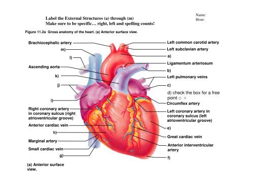
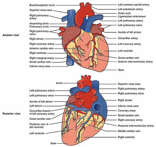
40 label the anterior view of the human heart
Human Heart - Diagram and Anatomy of the Heart - Innerbody The heart functions by pumping blood both to the lungs and to the systems of the body. To prevent blood from flowing backwards or "regurgitating" back into the heart, a system of one-way valves are present in the heart. The heart valves can be broken down into two types: atrioventricular and semilunar valves. Atrioventricular valves. The atrioventricular (AV) valves are located in the middle of the heart between the atria and ventricles and only allow blood to flow from the atria into ... Heart Labeling Quiz: How Much You Know About Heart Labeling? Here is a Heart labeling quiz for you. The human heart is a vital organ for every human. The more healthy your heart is, the longer the chances you have of surviving, so you better take care of it. Take the following quiz to know how much you know about your heart. Questions and Answers 1. What is #1? 2. What is #2? 3. What is #3? 4. What is #4?
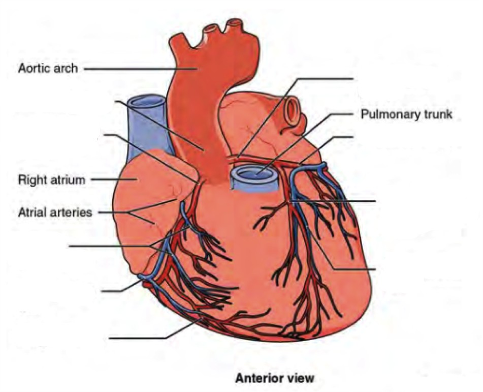
Anterior External View of Heart Labeling Diagram - Quizlet Anterior External View of Heart Labeling Diagram | Quizlet Anterior External View of Heart Labeling STUDY Learn Write Test PLAY Match Created by randajsmith Terms in this set (14) Superior Vena Cava ... Superior Vena Cava (SVC) ... Right Pulmonary Artery ... Right Pulmonary Veins ... Right Atrium ... Right Coronary Artery ... Right Ventricle ...

Label the anterior view of the human heart
1: Anatomy of the Heart (Anterior View): The figure illustrates the ... Atrial fibrillation (AF) is the most common cardiac arrhythmia in United States. The most popular treatment for AF is a percutaneous procedure called catheter ablation. Current AF ablation... Chapter 27 - Heart Anatomy - BIO 140 - Human Biology I - Textbook ... The dorsal surface of the heart lies near the bodies of the vertebrae, and its anterior surface sits deep to the sternum and costal cartilages. The great veins, the superior and inferior venae cavae, and the great arteries, the aorta and pulmonary trunk, are attached to the superior surface of the heart, called the base. PDF The Cardiovascular System - Pearson of the heart." The endocardium (en″do-kar′de-um) is a thin, glistening sheet of endothelium that lines the heart chambers. It is continuous with the linings of the blood vessels leaving and entering the heart. (Figure 11.3 shows two views of the heart—an exter-nal anterior view and a frontal section. As the ana-
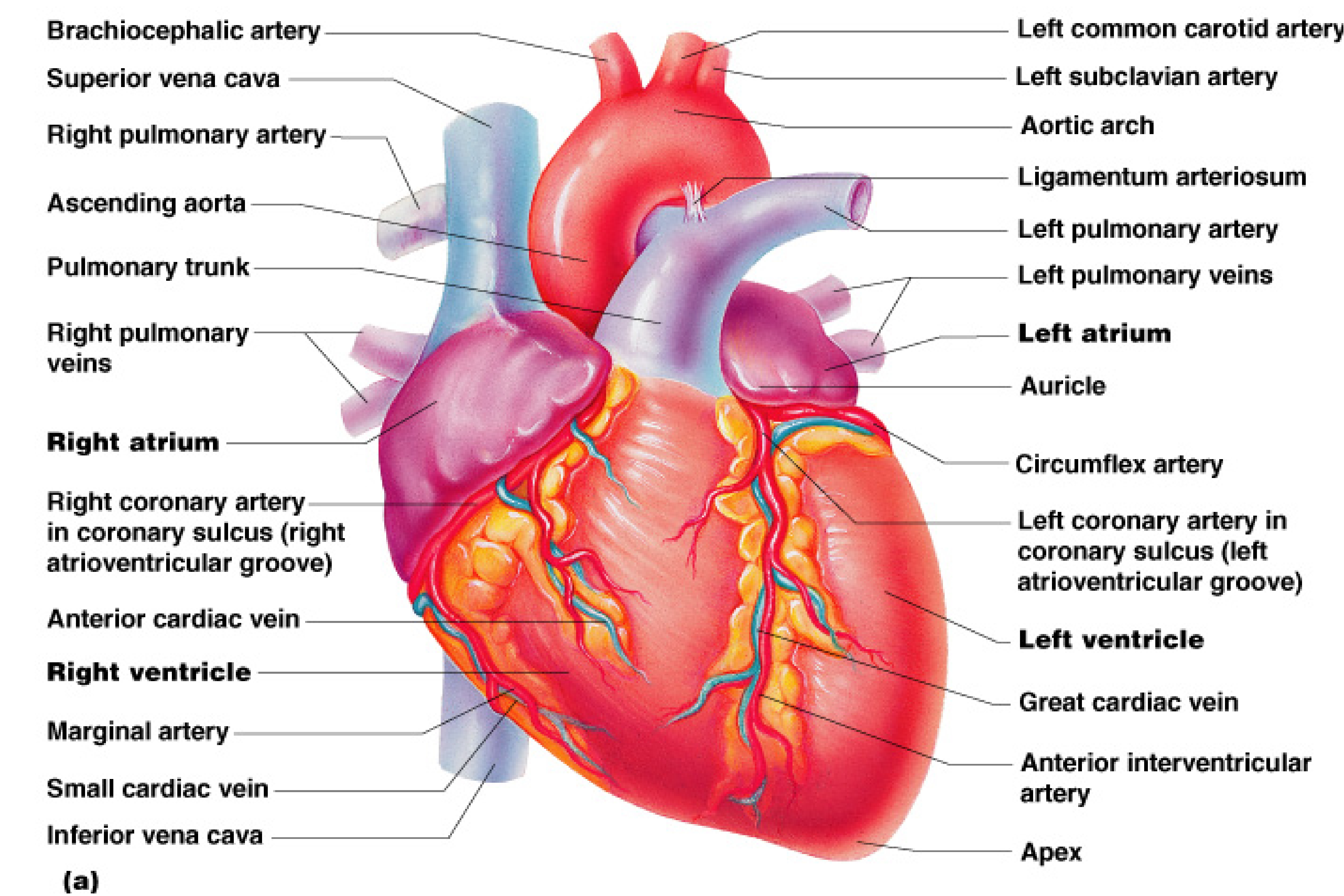
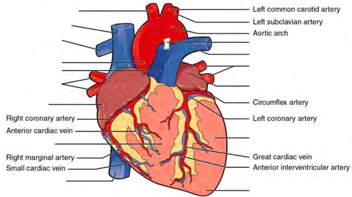
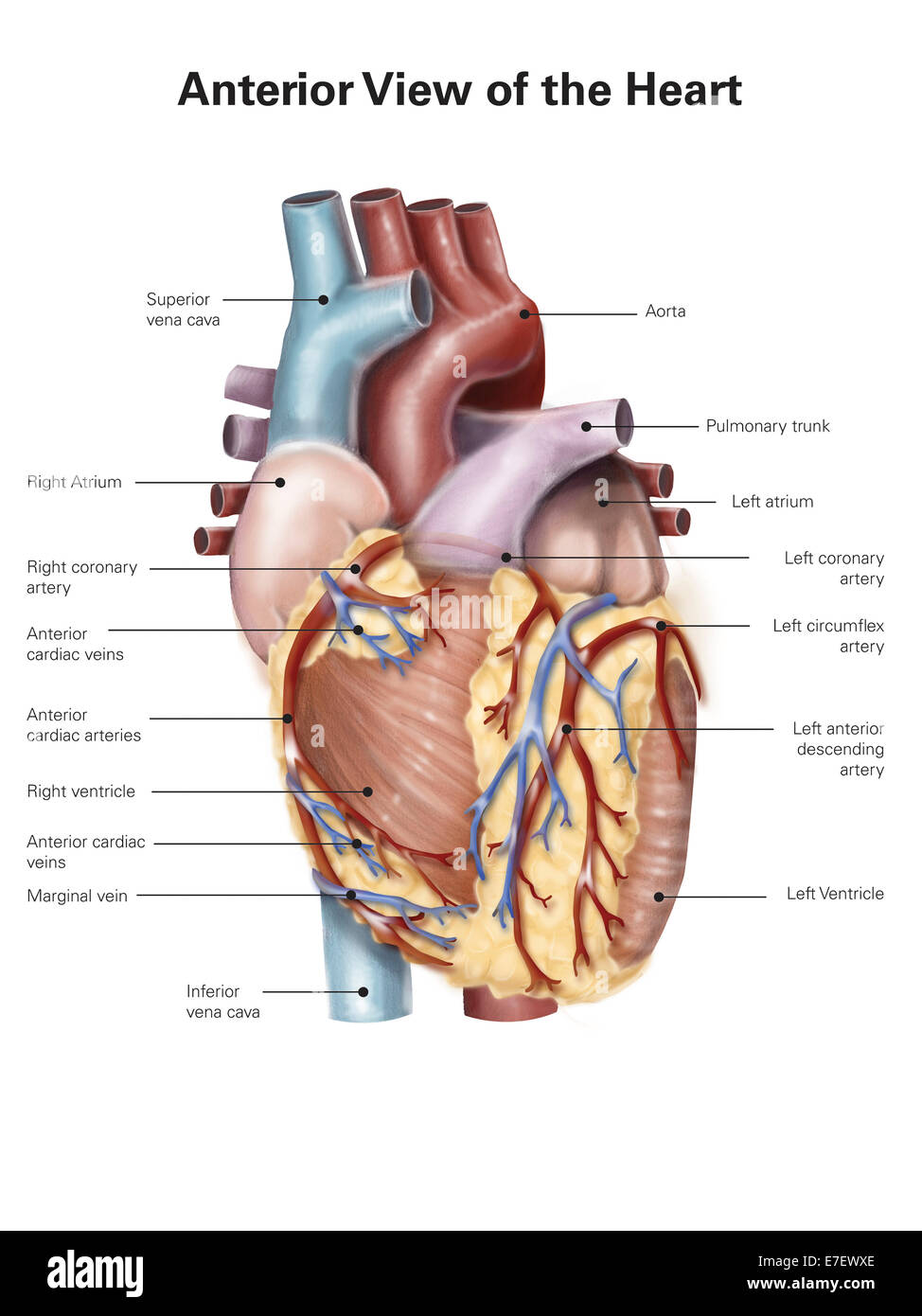
Label the anterior view of the human heart. Heart Labeling anterior view Diagram - Quizlet Heart Labeling anterior view Diagram | Quizlet Heart Labeling anterior view STUDY Learn Write Test PLAY Match + − Created by Meghan12th PLUS Terms in this set (26) brachiocephalic trunk ... left common carotid artery ... superior vena cava ... aortic arch ... liigamentum arteriosum ... right pulmonary artery ... amending aorta ... [Solved] labeling | Course Hero Heart 1. An anterior view of the heart is shown here. Match each structure listed with the correct letter in the figure. 1. right atrium B. brachiocephali... Show more ... Show more labeling Biology Science Anatomy Comments (2) Answer & Explanation Solved by verified expert All tutors are evaluated by Course Hero as an expert in their subject area. diagram of the heart unlabeled Heart - Anterior Gross Anatomy . anatomy heart anterior gross quiz external purposegames game. Draw A Sectional View Of Human Heart And Label, On It: - CBSE Class 10 ask.learncbse.in. cbse. System anatomy circulatory female femoral aorta arteries artery heart labeled major iliac popliteal abdominal structures aortic arch ... Anatomy Tutorial - Anterior | Atlas of Human Cardiac Anatomy This illustration demonstrates an anterior view of the thoracic cavity, highlighting the position of the heart in relationship to the ribs and diaphragm. The right atrium, right ventricle, and a small portion of the left ventricle are visible from this aspect. Note that in the majority of cases, 2/3 of the heart is positioned to the left of ...
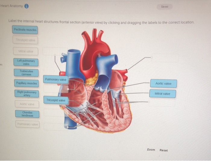
Solved Label this frontal section of the human heart by - Chegg Label this frontal section of the human heart by clicking and dragging the labels to the correct location. The arrows indicate direction of blood flow Right attium Aortic valve Lett pulmonary Pulmonary Superior vena Left ventricle artery valve Mitral valve Right pulmonary Pulmonary Tricuspid Inferior vena veis Right ventricle Loft atrium trunk valve cava Heart chambers and associated great vessels - Anatomy The right ventricle forms most of the anterior surface of the heart, while the left ventricle forms the heart apex. Two grooves on the heart surface indicate the boundaries of its four chambers and carry the blood vessels supplying the myocardium. Surfaces and Borders of the Heart - TeachMeAnatomy In its typical anatomical orientation, the heart has 5 surfaces, formed by different internal divisions of the heart: Anterior (or sternocostal) - Right ventricle. Posterior (or base) - Left atrium. Inferior (or diaphragmatic) - Left and right ventricles. Right pulmonary - Right atrium. Left pulmonary - Left ventricle. Heart anatomy: Structure, valves, coronary vessels - Kenhub Heart anatomy. The heart has five surfaces: base (posterior), diaphragmatic (inferior), sternocostal (anterior), and left and right pulmonary surfaces. It also has several margins: right, left, superior, and inferior: The right margin is the small section of the right atrium that extends between the superior and inferior vena cava .
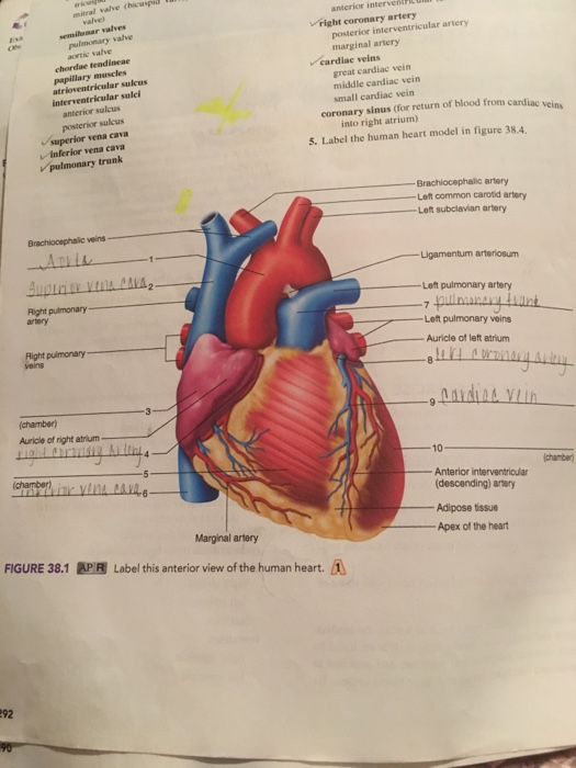
Anterior cardiac veins: Location, anatomy, function - Kenhub Anatomy and course. The 3-5 anterior cardiac veins emerge on the anterior surface of the right ventricle. They course horizontally across the surface of the ventricle, located deep to the epicardial layer of the heart.The anterior cardiac veins reach the atrioventricular groove, where they pass deep to the right coronary artery and empty directly into the right atrium. Heart - Collection Page | AnatomyTOOL A video of the combined view of the aortic and mitral valves in a cadaver heart that is beating in a laboratory set-up by the University of Minnesota. Several other videos of the combined views of beating aortic and mitral valves are on the Atlas page on the aortic valve as seen from the ventricle , see hearts: 416, 369, 235, (222), 136, 126-mitral and 53-1. Heart: illustrated anatomy - e-Anatomy - IMAIOS This interactive atlas of human heart anatomy is based on medical illustrations and cadaver photography. The user can show or hide the anatomical labels which provide a useful tool to create illustrations perfectly adapted for teaching. Anatomy of the heart: anatomical illustrations and structures, 3D model and photographs of dissection. Lab Report 38 Figures 38.1, 38.2, and 38.3.pdf - Course Hero Figure 38.1- Label this anterior view of the human heart. 1.) Aorta 2.) Left pulmonary artery 3.) Left pulmonary veins 4.) Left atrium 5.) Left ventricle 6.) Apex 7.) Superior vena cava 8.) Right atrium 9.) Inferior vena cava 10.)
External anterior heart labeling Quiz
Heart Diagram with Labels and Detailed Explanation - BYJUS Well-Labelled Diagram of Heart. The heart is made up of four chambers: The upper two chambers of the heart are called auricles. The lower two chambers of the heart are called ventricles. The heart wall is made up of three layers: The outer layer of the heart wall is called epicardium. The middle layer of the heart wall is called myocardium. The inner layer of the heart wall is called endocardium.
Fluid Mechanics Fundamentals and Applications 3 rd Edition
Draw a sectional view of the human heart and label the aorta, right ... Verified by Toppr. The right ventricle is the chamber of the heart that pumps out deoxygenated blood. Pulmonary vein is the blood vessel that carries oxygenated blood from the heart to different body parts. The pulmonary artery carries deoxygenated blood from different parts of the body to the heart. Aorta is the largest artery in the body.
Pre-lab 7 – Human Anatomy Lab Manual
Anterior view of the human heart. - Getty Images View top-quality illustrations of Anterior View Of The Human Heart. Find premium, high-resolution illustrative art at Getty Images.
Heart Gross Anatomy Practice Quiz
Solved Label external features of the anterior view of the | Chegg.com Label external features of the anterior view of the human heart. 3.5 pts Anterior inter- ventricular artery Aorta Left auricle Left coronary artery Pulmonary trunk Right auricle Right coronary artery ( r MODE Aortic valve Ascending aorta Bicuspid valve Left ventricle < MORE Chordae tendinae Papillary muscle Trabeculae carne Tricuspid valve leaflet.
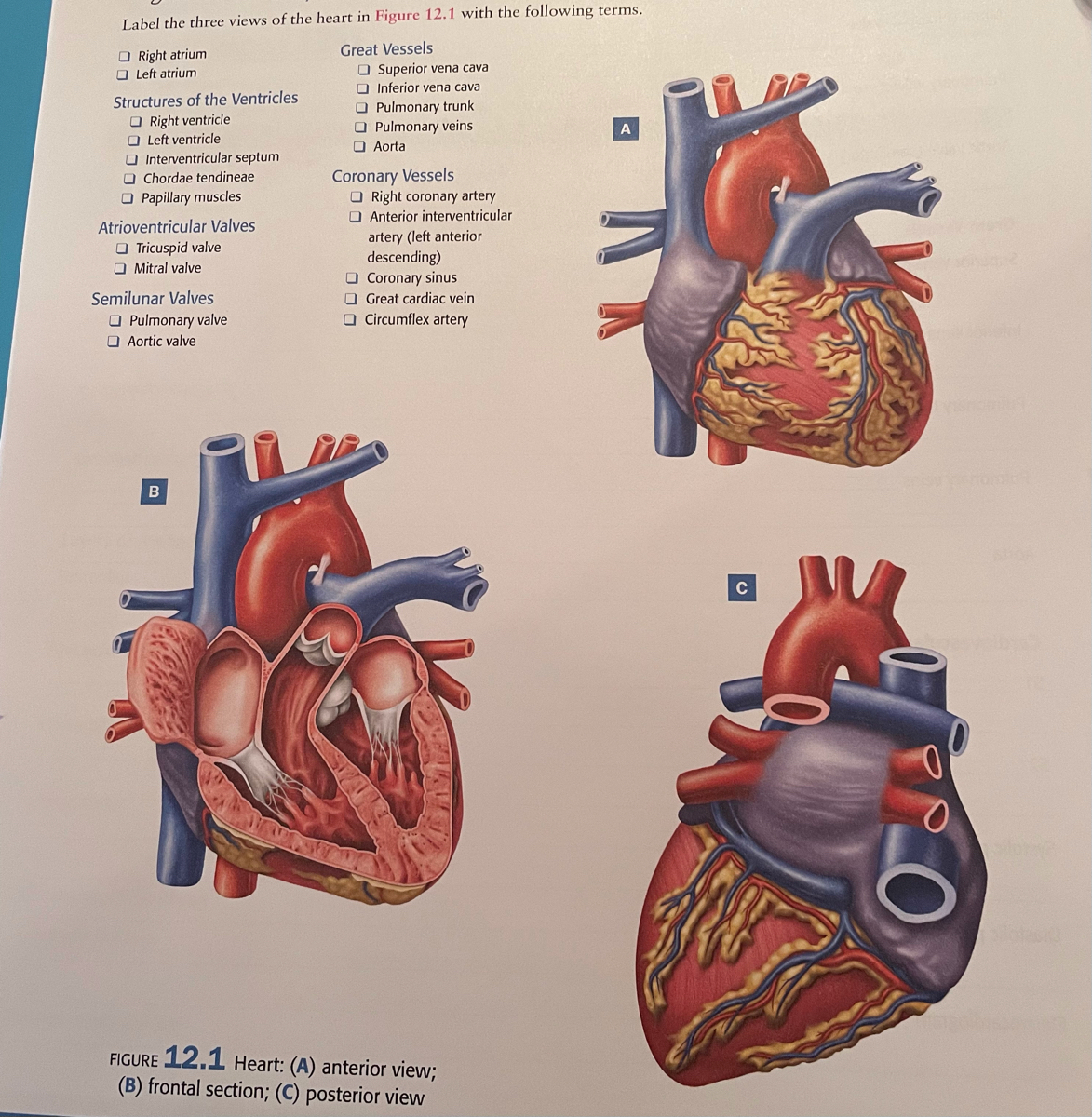
Answered: Label the three views of the heart in… | bartleby
Label the heart - Science Learning Hub Add to collection. In this interactive, you can label parts of the human heart. Drag and drop the text labels onto the boxes next to the diagram. Selecting or hovering over a box will highlight each area in the diagram. Right ventricle. Right atrium. Left atrium. Pulmonary artery. Left ventricle.
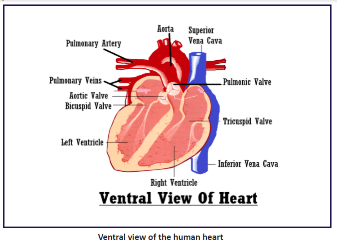
Sketch and label the ventral view of the human hea class 12 ...
Human Heart Anterior View Stock Illustration - Download Image Now - iStock iStock Human Heart Anterior View Stock Illustration - Download Image Now Download this Human Heart Anterior View vector illustration now. And search more of iStock's library of royalty-free vector art that features Diagram graphics available for quick and easy download. Product #: gm482925135 $ 33.00 iStock In stock
Sketch and label the ventral (anterior) view of human heart ...
Label the Heart Diagram Anterior view Quiz - PurposeGames.com This is an online quiz called Label the Heart Diagram Anterior view There is a printable worksheet available for download here so you can take the quiz with pen and paper. Your Skills & Rank Total Points 0 Get started! Today's Rank -- 0 Today 's Points One of us! Game Points 24 You need to get 100% to score the 24 points available Actions 2 favs

File:2008 Internal Anatomy of the HeartN.jpg - Wikimedia Commons
anterior heart Quiz - PurposeGames.com This is an online quiz called anterior heart There is a printable worksheet available for download here so you can take the quiz with pen and paper. From the quiz author quiz game to assist a&p students with anatomy of exterior heart. This quiz has tags. Click on the tags below to find other quizzes on the same subject. quiz heart label anterior

Chart of heart anterior view with parts name - vector image ...
Internal Structure of the Heart | Contemporary Health Issues The valves at the openings that lead to the pulmonary trunk and aorta are known generically as the pulmonary and the aortic valve. Structures of the Heart This anterior view of the heart shows the four chambers, the major vessels, and their early branches as well as the valves. Licenses and Attributions Previous Next
Anatomy of the Human Heart - Physiopedia
Anatomy of the Heart | Human heart anatomy, Gross anatomy, Human ... With exquisitely detailed illustrations, this 20" x 26" (51 x 66 cm) examination-room anatomy poster with grommets shows anterior, posterior and superior views of the heart. Anatomy of The Heart also depicts right and left ventricles, heart valves, blood circulation, cross section and anterior view of the heart with…

Anatomy And Physiology Heart Anatomy – Otosection
Human Heart (Anatomy): Diagram, Function, Chambers, Location in Body The heart is a muscular organ about the size of a fist, located just behind and slightly left of the breastbone. The heart pumps blood through the network of arteries and veins called the...
(230).jpg)
Heart Labeling Quiz: How Much You Know About Heart Labeling ...
PDF The Cardiovascular System - Pearson of the heart." The endocardium (en″do-kar′de-um) is a thin, glistening sheet of endothelium that lines the heart chambers. It is continuous with the linings of the blood vessels leaving and entering the heart. (Figure 11.3 shows two views of the heart—an exter-nal anterior view and a frontal section. As the ana-
Major Blood Vessels of the Heart | GetBodySmart
Chapter 27 - Heart Anatomy - BIO 140 - Human Biology I - Textbook ... The dorsal surface of the heart lies near the bodies of the vertebrae, and its anterior surface sits deep to the sternum and costal cartilages. The great veins, the superior and inferior venae cavae, and the great arteries, the aorta and pulmonary trunk, are attached to the superior surface of the heart, called the base.
Diagrams, quizzes and worksheets of the heart | Kenhub
1: Anatomy of the Heart (Anterior View): The figure illustrates the ... Atrial fibrillation (AF) is the most common cardiac arrhythmia in United States. The most popular treatment for AF is a percutaneous procedure called catheter ablation. Current AF ablation...

Anatomy of the Heart
Heart Anatomy | Anatomy and Physiology | | Course Hero

HEART, ILLUSTRATION Anatomy of the heart anterior view of a ...

Anterior View of Human Body Stock Illustration - Illustration ...
Real heart label the diagram of human heart animated real ...

Figure 35.1 Heart (ventral view) Diagram | Quizlet
Pre-lab 7 – Human Anatomy Lab Manual
Anterior view of the human heart Stock Photo - Alamy
Heart: Anatomy and Function

Heart chambers

Anatomy of the Heart | @thelibernation
What are the parts of the heart? - Quora

The Cardiovascular System: Blood and Sheep's Heart. - ppt ...

Heart Dissection Lab | Lt Anatomy Collection | ADI

Chapter 27 - Heart Anatomy - BIO 140 - Human Biology I ...

Anterior View of the Heart Diagram | Quizlet
is our

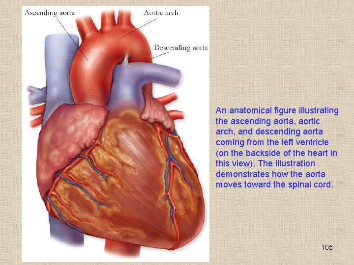
Anterior Surface View Of Heart - Spinal Cord - RR School Of ...
Solved Heart Anatomy Saved Label the internal heart | Chegg.com
Heart 3D Model: Top 5 Views
Heart ventricles: Anatomy, function and clinical aspects | Kenhub
The heart:
Draw a diagram of the front view of human heart and label any ...
Solved il valve ( haspid w ar waves pulmonary valve chordae ...
Heart Dissection Lab | Lt Anatomy Collection | ADI
Untitled
Post a Comment for "40 label the anterior view of the human heart"