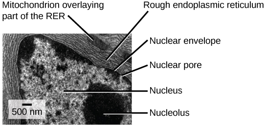
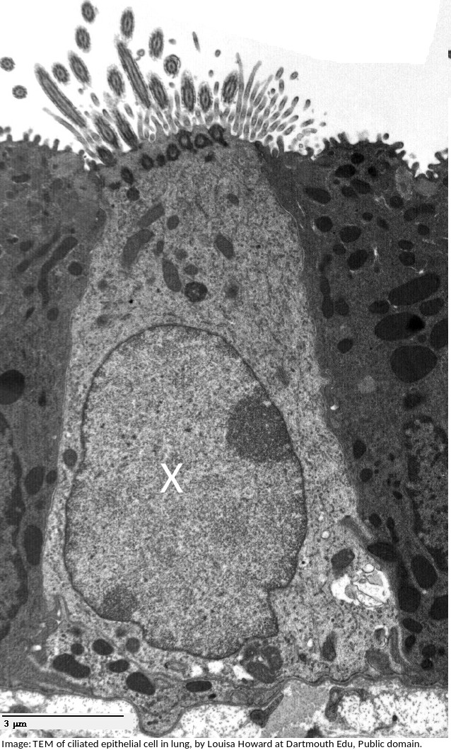
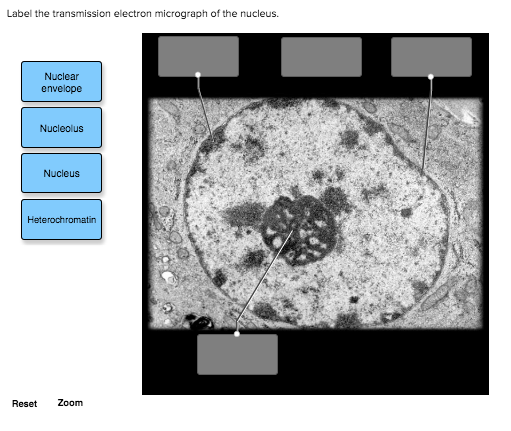
43 label the transmission electron micrograph of the nucleus.
Ultrastructure and nuclear architecture of telomeric chromatin revealed ... Here, we applied a correlative light and electron microscopy (CLEM) approach to study the structure of telomeres inside the nucleus at higher resolution. Telomeres were visualized by the expression of TRF1 or TRF2 fused in tandem to eGFP and the genetically encoded EM tag APEX2 (31,32). This allowed us to localize telomeres by confocal fluorescence microscopy and subsequently interrogate their ultrastructure and sub-nuclear organization by transmission electron microscopy (TEM), electron ... Category:Transmission electron microscopic images of cell nucleus Media in category "Transmission electron microscopic images of cell nucleus" ... Kernmembran nl.png 288 × 383; 58 KB. Kernmembran.png 288 × 383; 44 KB. Micrograph of a cell nucleus.png 200 × 199; 33 KB. NucleolusNCc.jpg 616 × 452; 120 KB ... Microscopic images of cell nucleus; Transmission electron microscopic images of organelles ...
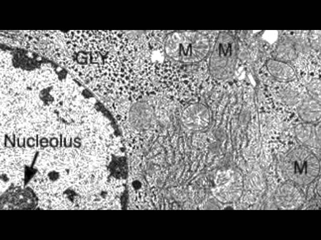
Plant Cell Nucleus Electron Micrograph : Cell And Organelles Dr Jastrow ... Us national institute of general medical a. The nucleus (plural = nuclei) figure 7.14 at left a transmission electron micrograph and at right a labeled diagram of a. Source: . Note the nucleus, nucleolus and condensed chromatin. Transmission electron micrograph (16,000x enlargement) through an infected root cell of a soybean plant.

Label the transmission electron micrograph of the nucleus.
A transmission electron micrograph of one of the 65 serial thin ... The centrosome of Dictyostelium discoideum is a nucleus-associated body consisting of an electron-dense, three-layered core surrounded by an amorphous matrix, the corona. Transmission electron microscopy of the bacterial nucleoid Abstract. Water-containing biological material cannot withstand the vacuum of the transmission electron microscope. The classical solution to this problem has been to dehydrate chemically fixed ... Label the transmission electron micrograph of the nucleus. Label the transmission electron micrograph of the cell. 0 Nucleus rences Mitochondrion Heterochromatin Peroxisome Vesicle ULAR bumit Click and drag each label into the correct category to indicate whether it pertains to the cytoplasm or the plasma...
Label the transmission electron micrograph of the nucleus.. Label This Transmission Electron Micrograph / Microscopy ... - Blogger Label This Transmission Electron Micrograph / Microscopy Innovations Transmission Electron Microscopy Tem. Label the transmission electron micrograph of the nucleus. Fluorescence microscopy in combination with tem and an ion beam analysis (iba, which allows the evaluation of the chemical elemental distribution) has allowed . Label the transmission electron micrograph of the cell. (d) a representative micrograph containing . Label This Transmission Electron Micrograph : TEM of chloroplast from ... Label the transmission electron micrograph of the nucleus. Molecular labeling for correlative microscopy: Subset of labeled images and transfer labels to the entire image corpus. Label this transmission electron micrograph of relaxed sarcomeres by clicking and dragging the labels to the correct location . Functional organization of the dorsal cochlear nucleus of the horseshoe ... Unique among mammals, the dorsal cochlear nucleus (DCN) of horseshoe bats consists of two functionally and anatomically distinct subdivisions: a laminated ventral portion that processes the frequency range below the constant frequency (CF) component of the echolocation signal and a nonlaminated dors … Electron microscopes - Cell structure - Edexcel - BBC Bitesize Electron microscopes use a beam of electrons instead of beams or rays of light. Living cells cannot be observed using an electron microscope because samples are placed in a vacuum. There are two ...
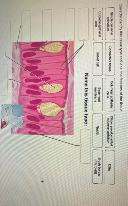
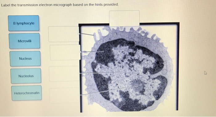
Solved Label the transmission electron micrograph of the | Chegg.com Transcribed image text: Label the transmission electron micrograph of the cell. 0 Nucleus rences Mitochondrion Heterochromatin Peroxisome Vesicle ULAR bumit Click and drag each label into the correct category to indicate whether it pertains to the cytoplasm or the plasma membrane. ICF Contacts the ECF Made of proteins and lipids Surrounds the cell Contains lon channels Organelles Fibers, tubulos, passages, and compartments Cytoskeleton Plasma Membrane Cytoplasm Correctly identify this tissue ... TEM of the Nucleus Unlike fluorescence microscopy, which relies upon the use of fluorescent probes to tag structures, TEM is capable of visualizing the structures themselves. The theoretical resolution of the transmission electron microscope is sufficient to resolve the molecular constituents of the individual nuclear compartments and structures. Light and electron microscope distribution of the NMDA ... - PubMed Light and electron microscope distribution of the NMDA receptor subunit NMDAR1 in the rat nervous system using a selective anti-peptide antibody ... nor did it label non-neuronal tissues. Immunostained vibratome sections of rat tissue showed labeling in many neurons in most structures in the brain, as well as in the cervical spinal cord, dorsal ... Solved Label the transmission electron micrograph of the - Chegg Expert Answer. Who are the experts? Experts are tested by Chegg as specialists in their subject area. We review their content and use your feedback to keep the quality high. 100% (23 ratings) Transcribed image text: Label the transmission electron micrograph of the nucleus. Nuclear envelope Nucleolus Nucleus Heterochromatin Reset Zoom.
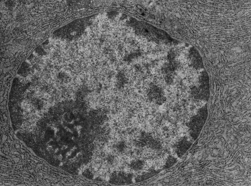
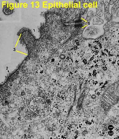
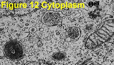
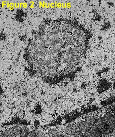
Cell Nucleus - function, structure, and under a microscope The double membrane system of the nuclear envelope (outer and inner membranes) is discovered by the transmission electron microscopic image below. You can see some gaps between the nuclear envelope; these are nuclear pores, like channels that allow transportation of molecules such as RNAs between the nucleus and the cytoplasm. Animal Cell Electron Microscope Labelled - Q14 Draw a large diagram of ... This is because of the way that the cell was sectioned (cut) before it was viewed on the transmission electron microscope. Source: hi-static.z-dn.net. Here is an electron micrograph of an animal cell with the labels superimposed: Animal cell electron micrograph labelling. The cell membrane is what controls the entry and exit of any substances ... Electron Micrographs** Electron Micrographs** Below is a collection of electron micrographs with labelled subcellular structures that you should be able to identify. Also, be sure to observe any electron micrographs which are made available in the laboratory by the instructor. ... Figure 1 Micrograph of a nucleus. 1. Heterochromatin 2. Euchromatin 3. Nucleolus 4 ... anatomy 10.png - Label the transmission electron micrograph of the ... anatomy 10.png - Label the transmission electron micrograph of the. anatomy 10.png - Label the transmission electron micrograph... School Utah Valley University; Course Title ZOOL 1090; Uploaded By emileeroylance19. Pages 1 Ratings 67% (3) 2 out of 3 people found this document helpful;
The increased osteoid in bone from hypocalcemic rats contains ...
Label-free three-dimensional imaging of cell nucleus using third ... The U.S. Department of Energy's Office of Scientific and Technical Information
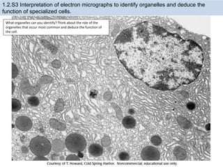
1.2 Obj Notes and Practice
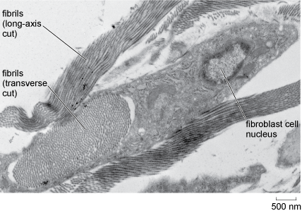
Transmission electron microscopy techniques - University of Otago The cytoplasm is full of mitochondria, lipid droplets, transparent vesicles, and an MII nucleus. Correlative TEM. Breast cancer cells by Sharon Lequeuex, read more below. Correlative microscopy involves using multiple microscope systems to observe the same specimen, most commonly light and transmission electron microscopy.

1.1 Cell structure | Cells as the basic units of life | Siyavula
Bio101 - Ch 6 HW Flashcards - Quizlet Tour of an Animal Cell: Part A. Drag the labels on the left onto the diagram of the animal cell to correctly identify the function performed by each cellular structure. a. smooth ER- synthesizes lipids. b. nucleolus- assembles ribosomes. c. defines cell shape. d. rough ER- produces secretory proteins.

Light and transmission electron micrographs of arabino- (1-4 ...
Electron Micrographs of Cell Organelles | Zoology The Electron Micrograph of Nucleus: This is an electron micrograph of nucleus. (Fig. 17 & 18): (1) Nucleus was discovered by Brown (1831). (2) It is a characteristic entity of almost all eukaryotic cells except mammalian RBCs. (3) The nucleus is generally one but may also be two, four or many.
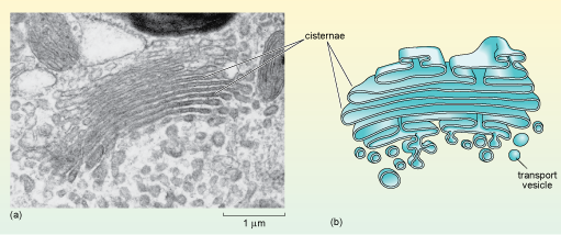
Biology, The Cell, Cell Structure, The Endomembrane System ...
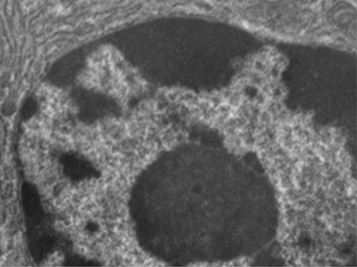
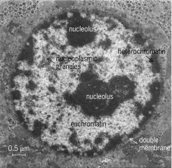
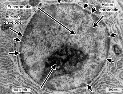
The Cell: The Histology Guide - University of Leeds This picture shows an electron micrograph of a nucleus. The short white arrows are pointing to nuclear pores. Note the appearance of eu- and heterochromatin, and the nucleolus. Heterochromatin stains more densely than euchromatin, but they are both forms of chromatin. Chromatin is the name for the diffuse granular mass of DNA found in ...

Cell Structure & Mitosis Visual Lab - ppt download
Labeling the Cell Flashcards - Quizlet Label the transmission electron micrograph of the nucleus. membrane bound organelles golgi apparatus, mitochondrion, lysosome, peroxisome, rough endoplasmic reticulum nonmembrane bound organelles ribosomes, centrosome, proteasomes cytoskeleton includes microfilaments, intermediate filaments, microtubules Identify the highlighted structures
A tour of the cell: View as single page
Transmission Electron Microscope (With Diagram) The specimen to be observed is placed on a copper mesh grid. Finally, the electrons are focused by an electromagnetic projector lens (instead of an ocular lens as in a light microscope) on a screen or photographic plate. The final image in a TEM is known as transmission electron micrograph.
Unique Characteristics of Eukaryotic Cells | Microbiology ...
Light and Electron Microscopy Study of Glycogen Synthase Kinase-3β in ... Electron microscopy images of GSK3β labeling in the piriform cortex. A) Partial image of a neuron (n) with an adjacent oligodendrocyte (o). The cytoplasm of the neuron presents abundant and strongly labeled rough endoplasmic reticulum (outlined arrows) and free ribosomes (outlined stars).
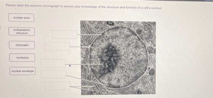
Solved Please label the electron micrograph to assess your ...
(Get Answer) - Electron micrograph of a plant cell with chloroplasts in ... (b) Electron micrograph of a white blood cell with mitochondria, a Golgi apparatus, and extensive rough endoplasmic reticulum in the cytoplasm around the large nucleus containing dark stained chromatin. 5. When a transmission electron microscope is used, cells are usually studied using electron micrographs, photographs taken of the image seen ...

Solved Mitochondrion Nucleus Vesicle Peroxisome | Chegg.com
PDF Identifying Organelles from an Electron Micrograph The electron micrograph displayed below illustrates many of the plant cell characteristics discussed The cell wall, large central vacuole and chloroplasts are clearly visible Also visible is the clearly defined nucleus containing chromatin Nucleus Chromatin The vacuole in this mature plant cell from a leaf is large, and occupies about 80% of

TEM of corneal buttons from patients with Fuchs' dystrophy ...
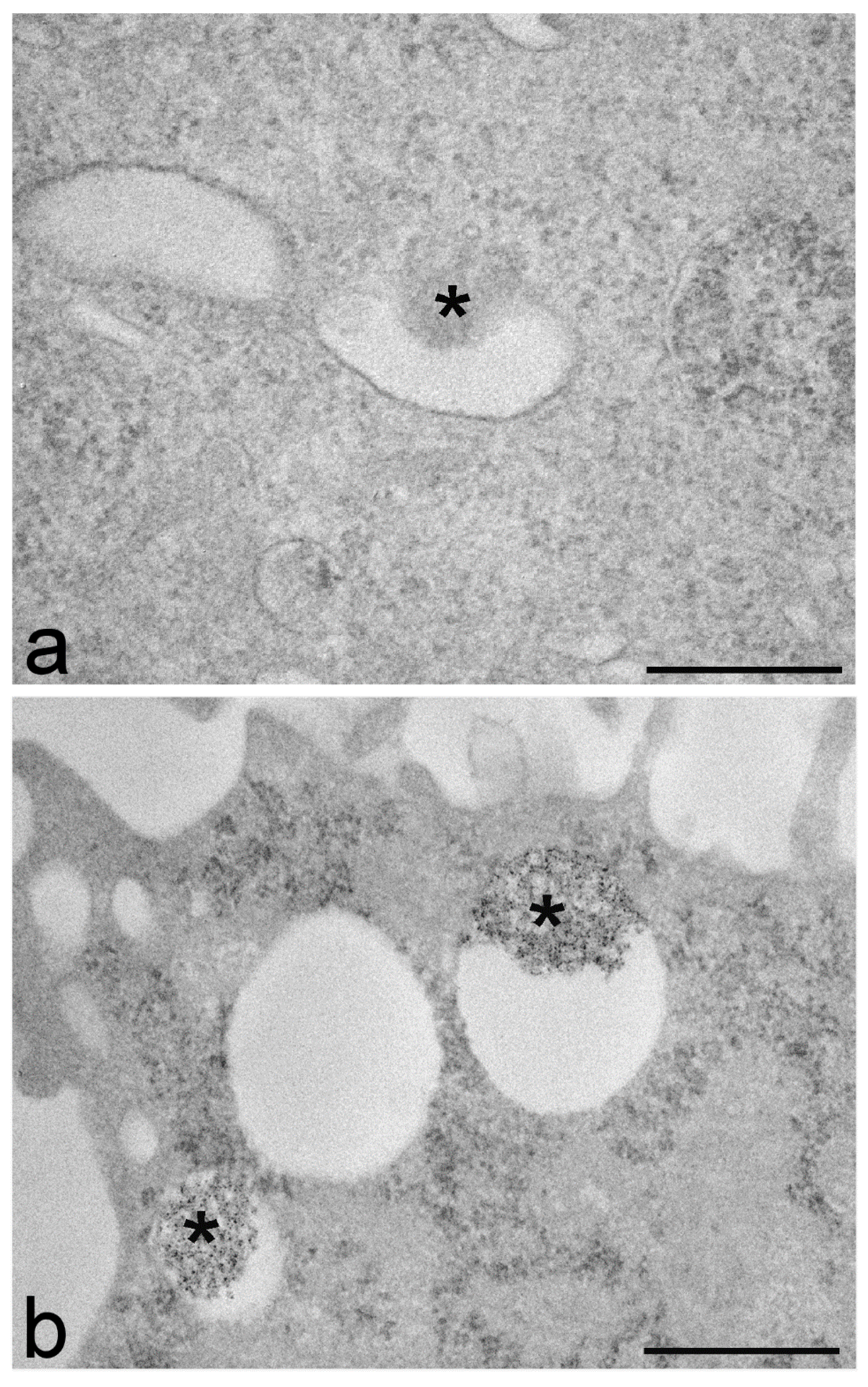
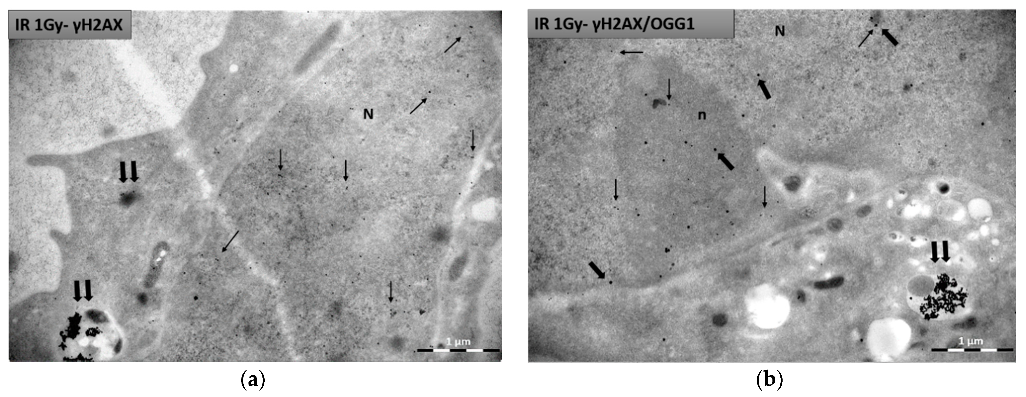
Metal-Tagging Transmission Electron Microscopy and Immunogold Labeling ... A scheme illustrating the labeling of cells infected with an IAV-MT recombinant virus. (a) The MT tag is fused with the PB2 subunit of the viral polymerase in vRNPs.(b, c) Scheme of METTEM (metal-tagging transmission electron microscopy) and silver enhancement .(b) In cells infected with the recombinant virus and incubated with gold salt, the MT tag builds a nanoparticle of ~1 nm.

Structure and Functions of the Dentin-Pulp Complex | Pocket ...
Label the transmission electron micrograph of the nucleus. Label the transmission electron micrograph of the cell. 0 Nucleus rences Mitochondrion Heterochromatin Peroxisome Vesicle ULAR bumit Click and drag each label into the correct category to indicate whether it pertains to the cytoplasm or the plasma...
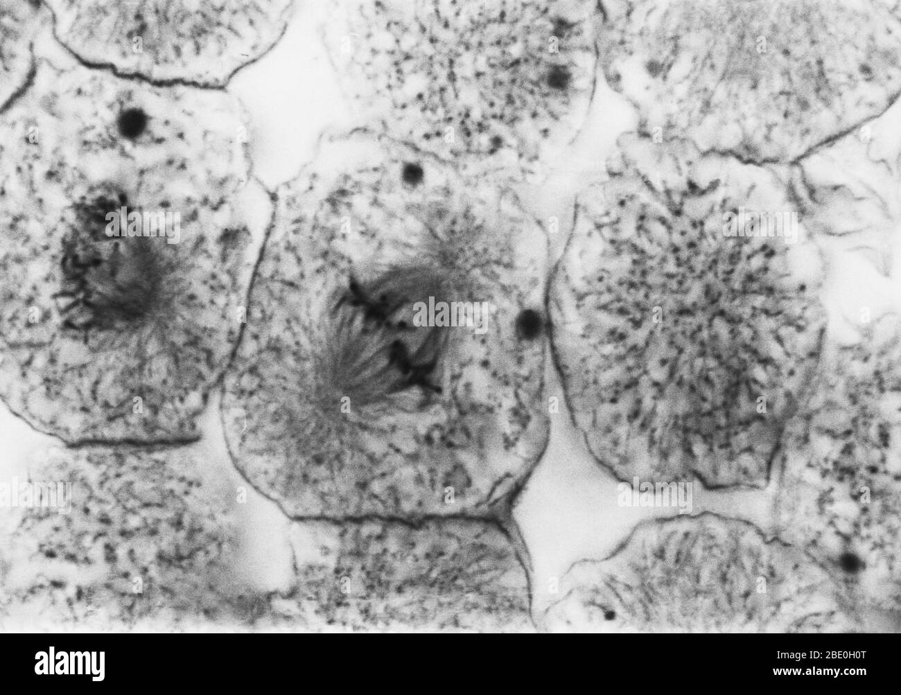
Metaphase hi-res stock photography and images - Alamy
Transmission electron microscopy of the bacterial nucleoid Abstract. Water-containing biological material cannot withstand the vacuum of the transmission electron microscope. The classical solution to this problem has been to dehydrate chemically fixed ...
Nucleus and nucleolus, TEM stock photo. Image of cytology ...
A transmission electron micrograph of one of the 65 serial thin ... The centrosome of Dictyostelium discoideum is a nucleus-associated body consisting of an electron-dense, three-layered core surrounded by an amorphous matrix, the corona.
Electron Micrographs
Solved Label the transmission electron micrograph of the ...
IJMS | Free Full-Text | Transmission Electron Microscopy as a ...
Ultrastructure of cells 1.2
Electron Micrographs

A Transmission electron micrograph (TEM) of a transverse ...
A tour of the cell: View as single page
What is a diagram of a plant and animal cell under an ...

Unique Characteristics of Eukaryotic Cells-Simplified ...
Cells (2.1, 2.2, 2.3, 2.4 & 2.5) Flashcards | Quizlet
IJMS | Free Full-Text | Visualization of Chromatin in the ...

4.9: Eukaryotic Cells - Mitochondria - Biology LibreTexts
Solved Label the transmission electron micrograph of the ...
Botrytis tulipae conidium: A. Transmission electron ...

A to 3D. Transmission electron microscopy (after 6 months). A ...
Histology and cytochemistry of interactions between plants ...
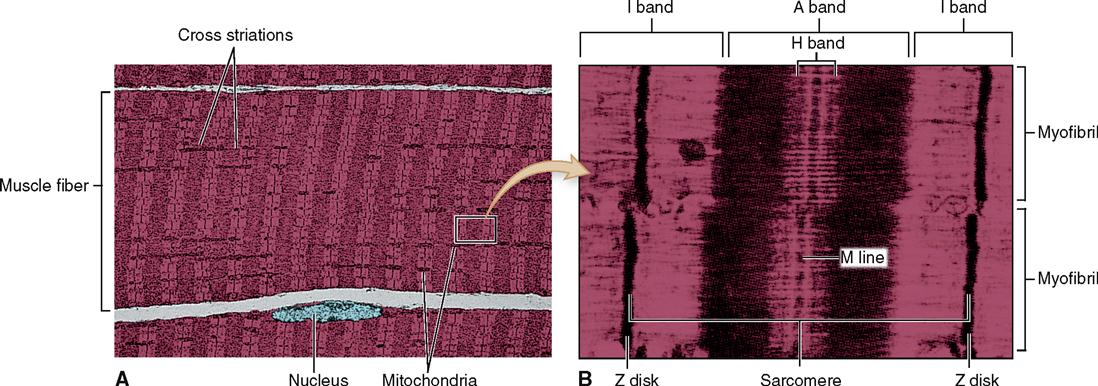
Physiology of the Muscular System | Basicmedical Key

Labeling the Cell Flashcards | Quizlet
Nanomaterials | Free Full-Text | A Guide for Using ...
Solved Label the transmission electron micrograph based on ...

DP Topic 1.1 / 1.2 | Biology - Quizizz

2.3.3 Identify structures from electron micrographs of liver ...

Transmission electron micrograph of an animal cell - Stock ...

Proteinuria induces tubular cell turnover: A potential ...
Electron Micrographs
2.3.3 Identify structures from electron micrographs of liver ...
The Cell: The Histology Guide

ELECTRON MICROSCOPY — Columbia Nano Initiative
Electron Micrographs
Post a Comment for "43 label the transmission electron micrograph of the nucleus."