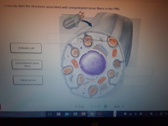
41 correctly label the structures associated with unmyelinated nerve fibers in the pns
Peripheral Nervous System | histology The CNS consists of the brain and the spinal cord, while the PNS is composed of nerves and groups of nerve cells (neurons), called ganglia. The nerves of the PNS carry sensory (afferent) inputs to the CNS and motor (efferent) output from the CNS to the skeletal and cardiac muscles and the smooth muscles of blood vessels, organs and glands. Which of the following are effectors a receptors b - Course Hero A) Receptors B) Stimuli C) Reflexes D) Glands E) Sense organs. 19) What is another name for the autonomic nervous system? A) Visceral sensory division B) Somatic sensory division C) Visceral motor division D) Somatic motor division E) Central nervous system. 20) Nerves are __________ of the nervous system. A) organs.
16.4 The Peripheral Nervous System - Concepts of Biology - 1st Canadian ... The peripheral nervous system (PNS) is the connection between the central nervous system and the rest of the body. The CNS is like the power plant of the nervous system. It creates the signals that control the functions of the body. The PNS is like the wires that go to individual houses. Without those "wires," the signals produced by the ...

Correctly label the structures associated with unmyelinated nerve fibers in the pns
Free Science Flashcards about ANP1040 Exam 4 - StudyStack Correctly label the structures, areas, and concentrations associated with a cell's electrical charge difference across its membrane. ... Spinal nerve, Perineum, Myelin, Epineuriun, Endoneurium, Myelinated nerve fiber, Unmyelinated nerve fiber: Which of the following structures is the richest in lipid content? -arachnoid mater -pia mater -white ... Muscle and Nervous Tissue Review Flashcards | Quizlet Terms in this set (50) Place the organizational level of muscle tissue in order, beginning with the entire muscle and ending with the smallest component. 1.) Muscle 2.) Fascicle 3.) Muscle Fiber 4.) Myofibril 5.) Myofilament Label the components of skeletal muscle. Label the connective tissue in the figure. lab 6 answers.docx - 201 LAB 6: NERVOUS SYSTEM With Answers... 216 ear models - label using the following terms: oval window, cochlea, cochlear nerve, vestibular nerve, tympanic cavity, round window, vestibule, semicircular canal, tympanic membrane, stapes, malleus, incus, inner ear, middle ear, external ear, auricle or pinna, external auditory canal, tympanic membrane 8 new 9 auricle or pinna external …
Correctly label the structures associated with unmyelinated nerve fibers in the pns. 16.1 Neurons and Glial Cells - Concepts of Biology - 1st Canadian Edition Figure 16.2. Nervous systems vary in structure and complexity. In (a) cnidarians, nerve cells form a decentralized nerve net. In (b) echinoderms, nerve cells are bundled into fibers called nerves. In animals exhibiting bilateral symmetry such as (c) planarians, neurons cluster into an anterior brain that processes information. Unit 4 Anatomy & Physiology Flashcards | Quizlet The Schwann cell's plasma membrane spirals repeatedly around the unmyelinated fiber as it does in a myelin sheath. false place the following in the order that an electrical impulse would travel beginning with the post-synaptic membrane 1. dendrite 2. soma 3. axon hillock 4. internode 5. node of rangier 6. terminal aborization 7. synaptic knobs Nervous Tissue - Characteristics, Structure, Function - BYJUS Nervous or the nerve tissue is the main tissue of our nervous system. It monitors and regulates the functions of the body. Nervous tissue consists of two cells: nerve cells or neurons and glial cells, which helps transmit nerve impulses and also provides nutrients to neurons. Brain, Spinal Cord, and nerves are composed of nervous tissue, they are specialized for being stimulated to transmit ... Ch 14 Flashcards | Chegg.com Describe the main steps in the regeneration of a nerve fiber. Describe a typical chemical synapse in reference to its microanatomy and the role of the neurotransmitters. Define the following terms and correctly match with either the CNS or PNS: ganglia, nerve, center (or nucleus), tract (or fascicle), and column.
Divisions of the Autonomic Nervous System | Anatomy and Physiology I These fibers are unmyelinated. (Note that the term "postganglionic neuron" may be used to describe the projection from a ganglion to the target. The problem with that usage is that the cell body is in the ganglion, and only the fiber is postganglionic. Typically, the term neuron applies to the entire cell.) Parasympathetic Nervous System Functions | Simply Psychology Nerve fibres of the PSNS arise within the central nervous system. The primary nerves involved are cranial nerves. Below are some of the main cranial nerves in the PSNS: Vagus nerve - approximately 75% of all the parasympathetic nerves are vagus nerves. These nerves have branches in many key organs such as the stomach, kidneys, bladder, and ... Fundamental the Nervous System an vous Ti - lake.k12.fl.us Functions and Divisions of the Nervous System conduction along unmyelinated fibers. 1. List the basic functions of the nervous system. ... The process called a nerve fiber 9. Formed by Schwann cells in the PNS 10. Clustered ribosomes and rough ER ... associated) structures described here. Write the correct terms in the answer blanks. 1 ... DiFiore's Atlas of Histology with Functional Correlations ... There is shortage of references in higher teaching institutions especially in newly opened institutions engaged in training of various Veterinary professionals in the country.
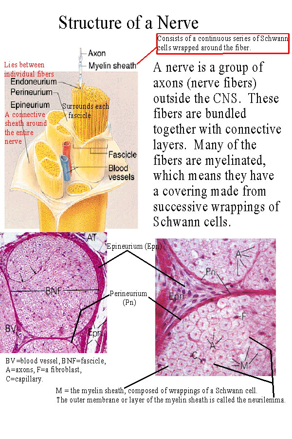
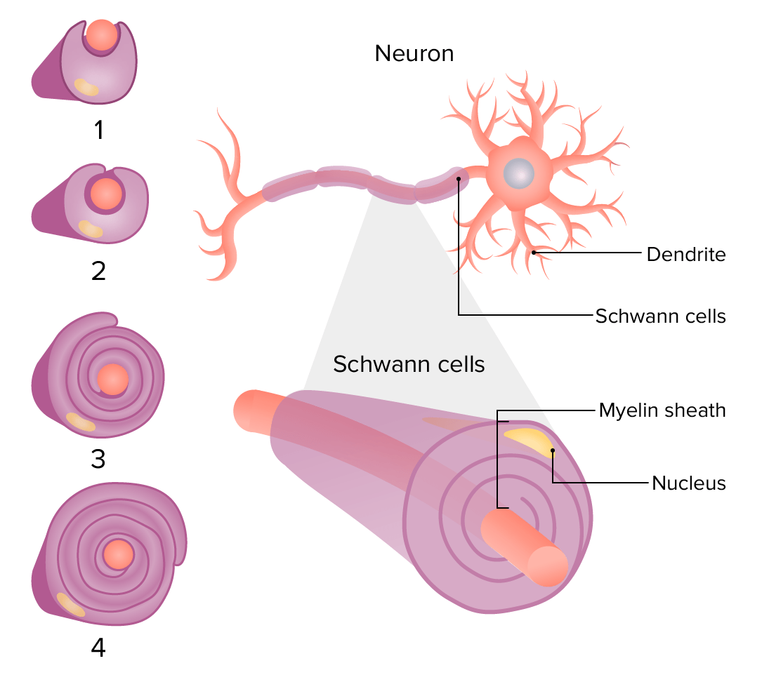
Be able to diagram and label a nerve cross section - Course Hero fasiculus (fascicle), epineurium, perineurium, endoneurium, neurolemmocyte, o Neurolemmocytes, also called Schwann cells, are associated with PNS axons. They are elongated, flattened cells that wrap around axons withinthe PNS, insulating the axon and forming a myelin sheath axon Anatomy, Central Nervous System - StatPearls - NCBI Bookshelf These regions are then broken down into 31 segments with 31 pairs of spinal nerves. There are 8 cervical nerves, 12 thoracic nerves, 5 lumbar nerves, 5 sacral nerves, and 1 coccygeal nerve. Each nerve exits the vertebral column passing through the intervertebral foramina and to its designated location in the body. A & P Exam 3 Nervous System Example | GraduateWay central nervous system contains brain and the spinal cord. PNS. peripheral nervous system consisting of nerves and ganglia that lie outside of the brain and spinal cord. somatic nervous system. the division of the PNS that provides the motor innervation of skeletal muscles; voluntary nervous (skeletal muscle) system. dermatomes. 23 some neurons are specialized to detect stimuli - Course Hero A Schwann cells wraps its plasma membrane around each individual fiber as it does with myelinatedfibers. An oligodendrocyte cells wraps its plasma membrane around each fiber as it does with myelinated fibers.Satellite cells cluster around each axon to form a pseudo-myelin sheath. A Schwann cell folds its plasma membrane around several fibers . 25.
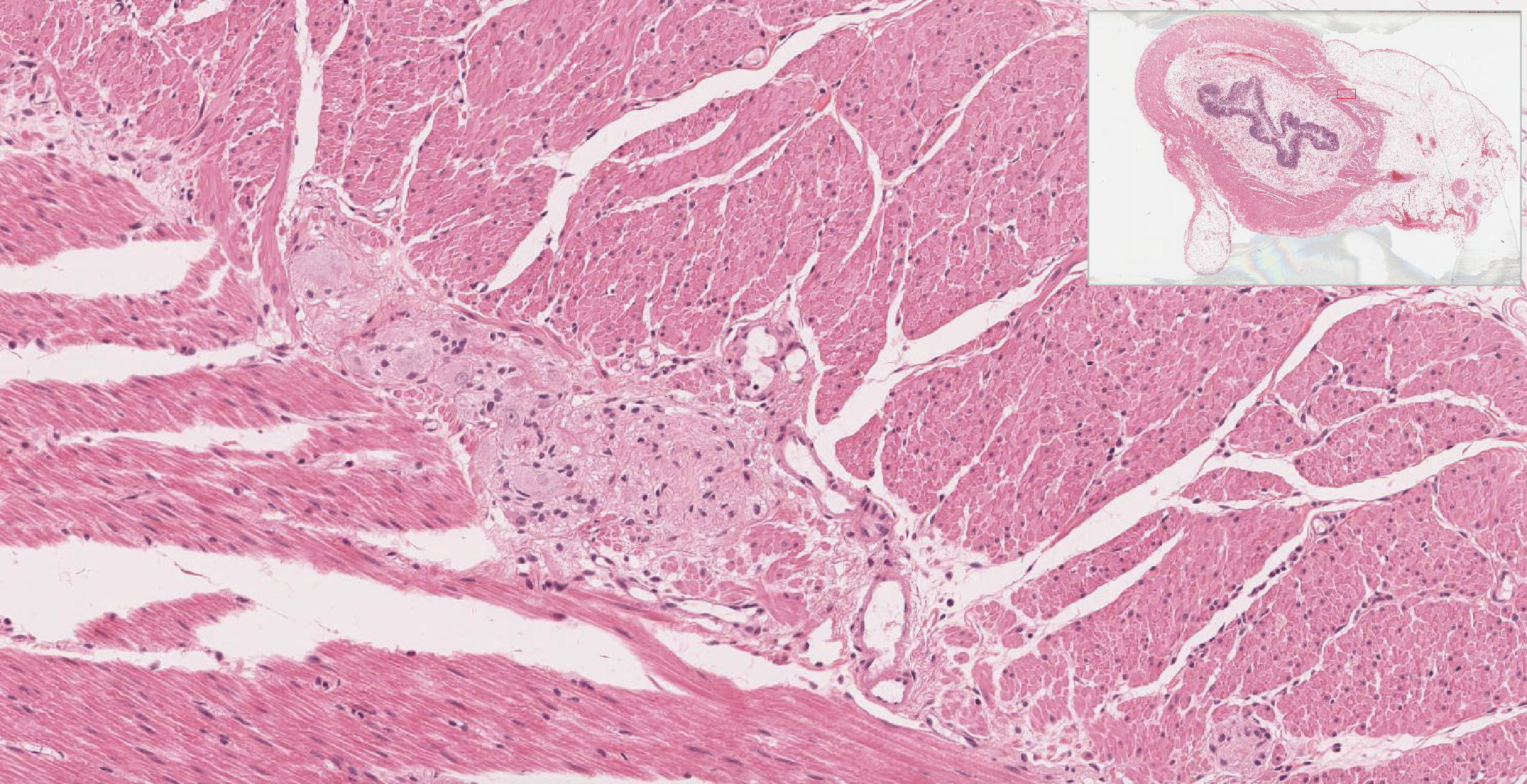
Histology of the Peripheral Nerves and Light Microscopy ...
Lab 1 Homework BIOL 320 Flashcards | Quizlet Correctly label the structures associated with unmyelinated nerve fibers in the PNS. Indicate the specified region of the diencephalon. Thalamus. Determine which general anatomic feature of the brain is illustrated in the figure. Sulcus. Determine which specific tract is depicted in the figure.
BIOL 237 Class Notes - The Spinal Cord and Spinal Nerves
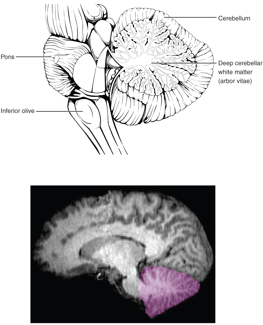
4. Neuroanatomy CNS.pdf - 4/25/2018 4. Neuroanatomy CNS 4.... - Course Hero ANSWER: Correct The cerebral hemispheres, which form the superior part of the brain, account for about 83% of total brain mass.Nearly the entire surface of the cerebral hemispheres is marked by elevated ridges called sulci. The cerebral hemispheres account for about 83% of total brain mass.

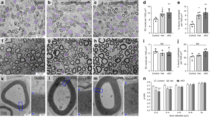
CNS Hypomyelination Disrupts Axonal Conduction and Behavior ...
Anatomy Midterm Lecture Flashcards | Quizlet • A membrane potential reading of +10 mV • Inactivated voltage-gated sodium channels • Open voltage-gated potassium channels Repolarization Label each phase of the action potential as identified by the highlighted region of each graph. Action potentials occur ____________________________. in the unmyelinated regions of an axon.
Frontiers | Biomaterials for Neural Tissue Engineering
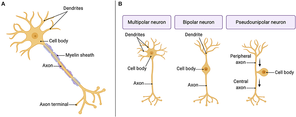
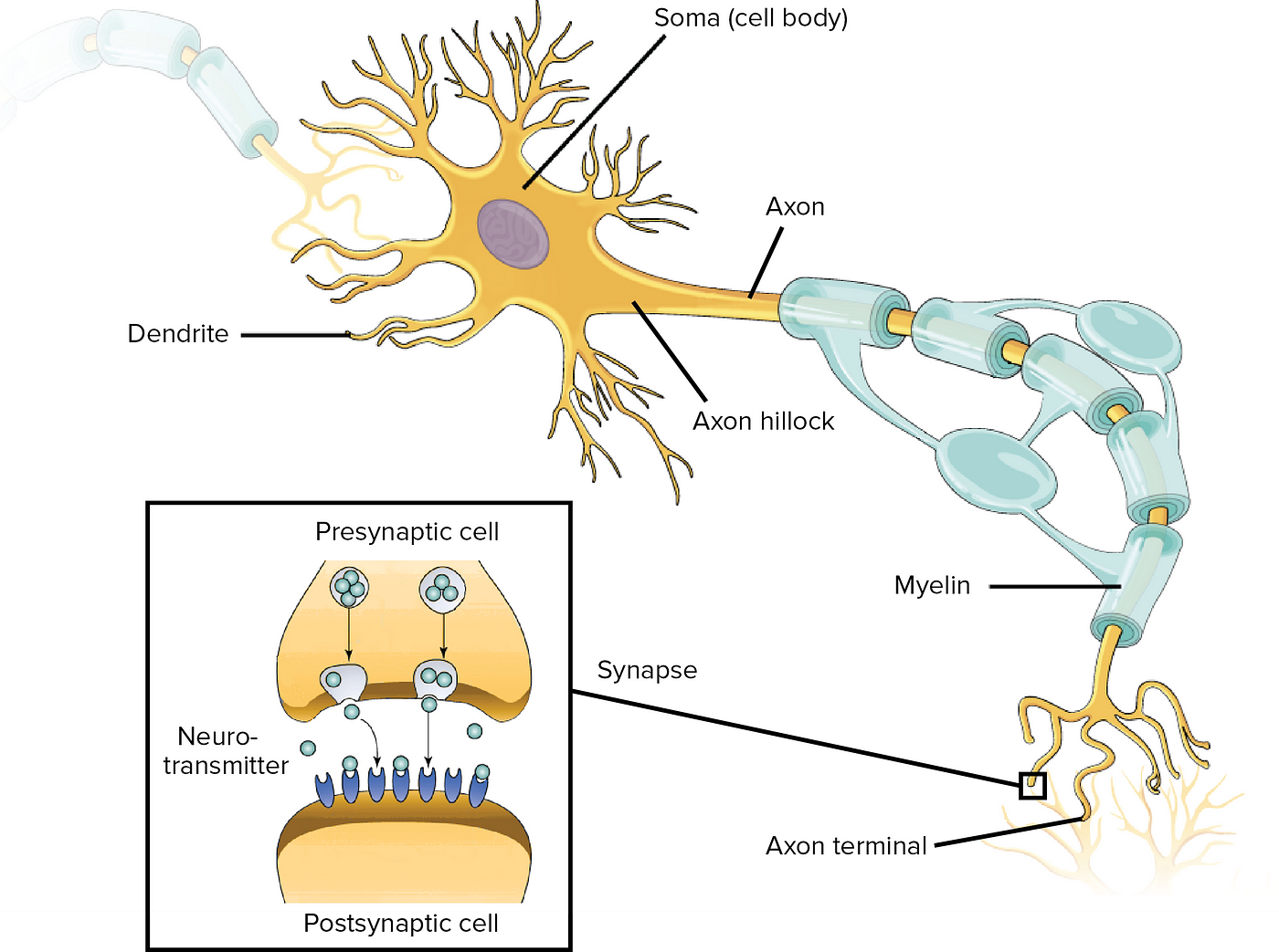
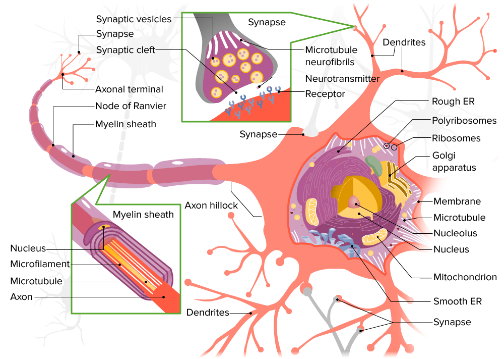
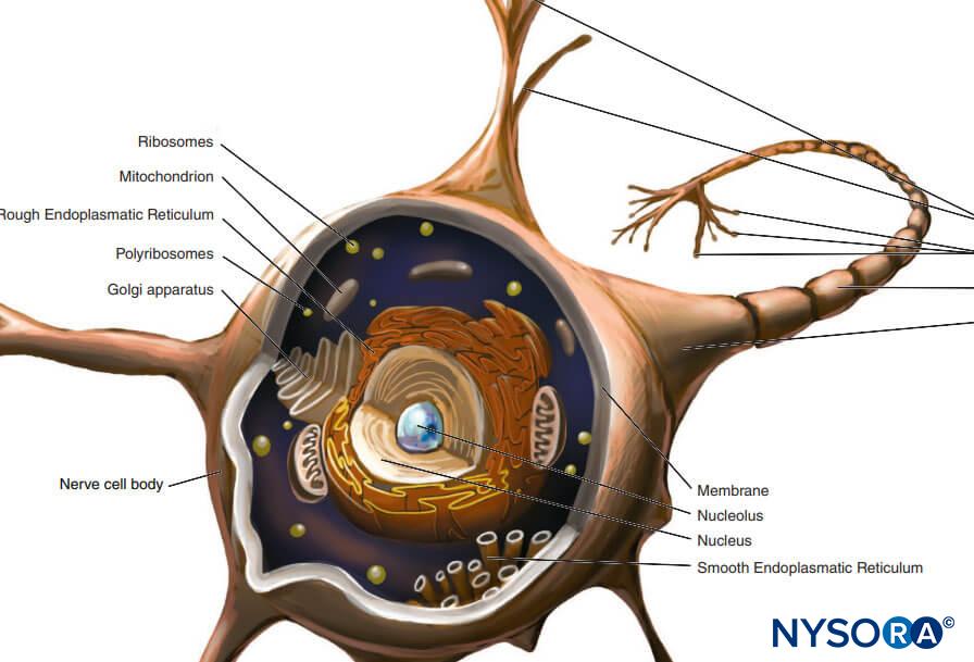
Nervous system: Structure, function and diagram | Kenhub Neurons, or nerve cell, are the main structural and functional units of the nervous system. Every neuron consists of a body (soma) and a number of processes (neurites). The nerve cell body contains the cellular organelles and is where neural impulses ( action potentials) are generated. The processes stem from the body, they connect neurons with ...

Central nervous system hypomyelination disrupts axonal ...
PDF Principles of Nerve Conduction Studies - AANEM the specific use described by the authors and are "off-label" (i.e. use not described on the product's label). ... motor NCS and NEE assess the motor nerve fibers of the PNS ... The motor and sensory axons composing the PNS may be myelinated or unmyelinated. The myelin does not coat the nerve fiber uniformly but, rather, in segments. ...

10.3 Muscle Fiber Excitation, Contraction, and Relaxation ...
Solved Place a single word into each sentence to make it | Chegg.com question: place a single word into each sentence to make it correct, describing the repair of a peripheral nerve fiber. lamina a damaged nerve fiber may regenerate if its soma is intact and some neurilemma remains. cannot soma when a nerve fiber is cut, the fiber distal to the injury survive. peripheral near the site of injury, the basal lamina …
Neuroanatomy for the Rest of Us. The basics of brain anatomy ...
Chapter 12 QS Anatomy (Nervous System) Flashcards | Quizlet Consists of the brain and spinal cord=central nervous system Includes cranial nerves, spinal nerves, and ganglia=peripheral nervous system When a neurotransmitter binds a protein channel, it opens and lets sodium diffuse down its concentration gradient. This is an example of a chemically gated sodium channel.

Anatomy Midterm Lecture Flashcards | Quizlet
Peripheral Nervous System: Spinal Nerves and Plexuses - Antranik The ventral rami in the thoracic region are known as the intercostal nerves . They run deep to the ribs and innervate the intercostal muscles and provide sensory input for the overlying horizontal strips of skin there as well as the abdominal wall muscles (motor) and skin (sensory).
PDF) The Role of Neurotropic B Vitamins in Nerve Regeneration
The Peripheral Nervous System | SEER Training Each bundle of nerve fibers is called a fasciculus and is surrounded by a layer of connective tissue called the perineurium. Within the fasciculus, each individual nerve fiber, with its myelin and neurilemma, is surrounded by connective tissue called the endoneurium. A nerve may also have blood vessels enclosed in its connective tissue wrappings.
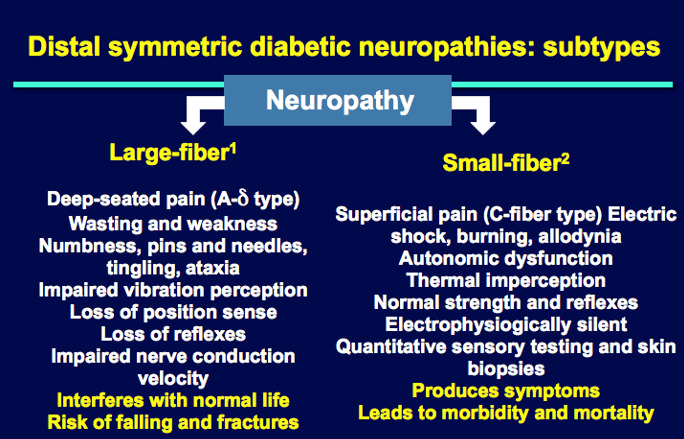
Diabetic Neuropathies - Endotext - NCBI Bookshelf
Action potential - Definition, Steps, Phases | Kenhub An action potential is defined as a sudden, fast, transitory, and propagating change of the resting membrane potential. Only neurons and muscle cells are capable of generating an action potential; that property is called the excitability. This article will discuss the definition, steps and phases of the action potential.

Focused Ultrasound (FUS) for Chronic Pain Management ...
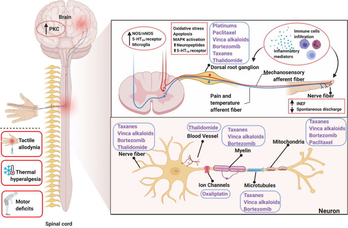
Dorsal root of spinal nerve - Wikipedia The dorsal root ganglia contain the pseudo-unipolar cell bodies of the nerve fibres which travel from the ganglia through the root into the spinal cord. The lateral division of the dorsal root contains lightly myelinated and unmyelinated fibres of small diameter [citation needed]. These carry pain and temperature sensation.

What Is Central Nervous System? Definition, Function & Parts
Solved CORRECTLY LABEL THE STRUCTURES ASSOCIATED WITH - Chegg Expert Answer. 100% (18 ratings) Answer: Correctly …. View the full answer. Transcribed image text: Correctly label the structures associated with unmyelinated nerve fibers in the PNS Schwann cel Unmyelinated nerve nbers Basal lamina 閰咇.
Nervous System: Histology | Concise Medical Knowledge
lab 6 answers.docx - 201 LAB 6: NERVOUS SYSTEM With Answers... 216 ear models - label using the following terms: oval window, cochlea, cochlear nerve, vestibular nerve, tympanic cavity, round window, vestibule, semicircular canal, tympanic membrane, stapes, malleus, incus, inner ear, middle ear, external ear, auricle or pinna, external auditory canal, tympanic membrane 8 new 9 auricle or pinna external …

Cranial nerves - Wikipedia
Muscle and Nervous Tissue Review Flashcards | Quizlet Terms in this set (50) Place the organizational level of muscle tissue in order, beginning with the entire muscle and ending with the smallest component. 1.) Muscle 2.) Fascicle 3.) Muscle Fiber 4.) Myofibril 5.) Myofilament Label the components of skeletal muscle. Label the connective tissue in the figure.
Nervous System: Histology | Concise Medical Knowledge
Free Science Flashcards about ANP1040 Exam 4 - StudyStack Correctly label the structures, areas, and concentrations associated with a cell's electrical charge difference across its membrane. ... Spinal nerve, Perineum, Myelin, Epineuriun, Endoneurium, Myelinated nerve fiber, Unmyelinated nerve fiber: Which of the following structures is the richest in lipid content? -arachnoid mater -pia mater -white ...

Histology of the Peripheral Nerves and Light Microscopy ...
Intro to NS & Nervous Tissue Ch11-Student handoutF15.pptx

Veterinary Neurobiology Courseware

What Is a Neuron? Diagrams, Types, Function, and More

51139882-C1CB-4A6C-ABC6-DB03F69453B6.jpeg - Correctly label ...
Peripheral Nervous System | histology

Applied Basic Sciences (Section 5) - Postgraduate Orthopaedics
Targeting strategies for oxaliplatin-induced peripheral ...
Full article: Axonal mRNA translation in neurological disorders
Structure and function of connective tissue (Chapter 20 ...
Nervous system: Structure, function and diagram | Kenhub

PDF) Myelin Fat Facts: An Overview of Lipids and Fatty Acid ...

Myelin Sheath Function & Type of Conduction | Schwann Cells ...
Limbic System - Physiopedia
DR. NAITIK D TRIVEDI & DR. UPAMA N. TRIVEDI

AHCDW8Notes10.pdf - 10. Award: 10.00 points Problems? Adjust ...
Distribution of nerve fibers and nerve-immune cell ...
Nervous System – Building a Medical Terminology Foundation
Histology of the Peripheral Nerves and Light Microscopy ...
Untitled

Nerve guide conduits for peripheral nerve injury repair: A ...
Myelinating Schwann cells ensheath multiple axons in the ...
Solved CORRECTLY LABEL THE STRUCTURES ASSOCIATED WITH | Chegg.com

Conservation and divergence of myelin proteome and ...

Specific gene expression in unmyelinated dorsal root ganglion ...

Exosomes as Neurological Nanosized Machines | ACS Nanoscience Au
Post a Comment for "41 correctly label the structures associated with unmyelinated nerve fibers in the pns"