42 label the photomicrograph of the sebaceous gland.
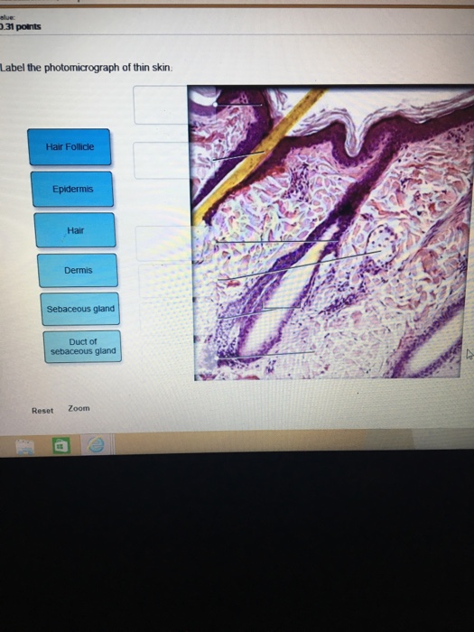
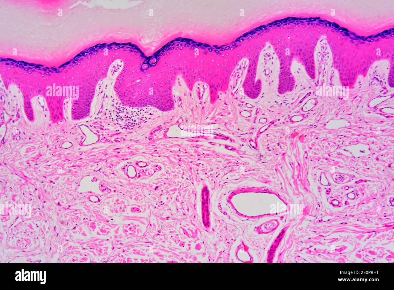
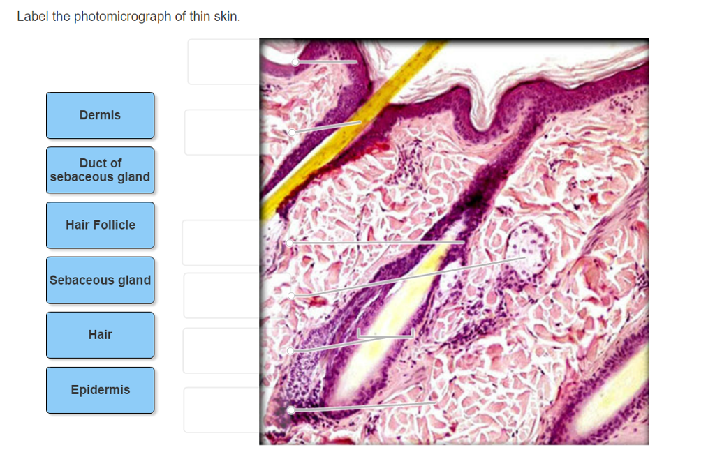
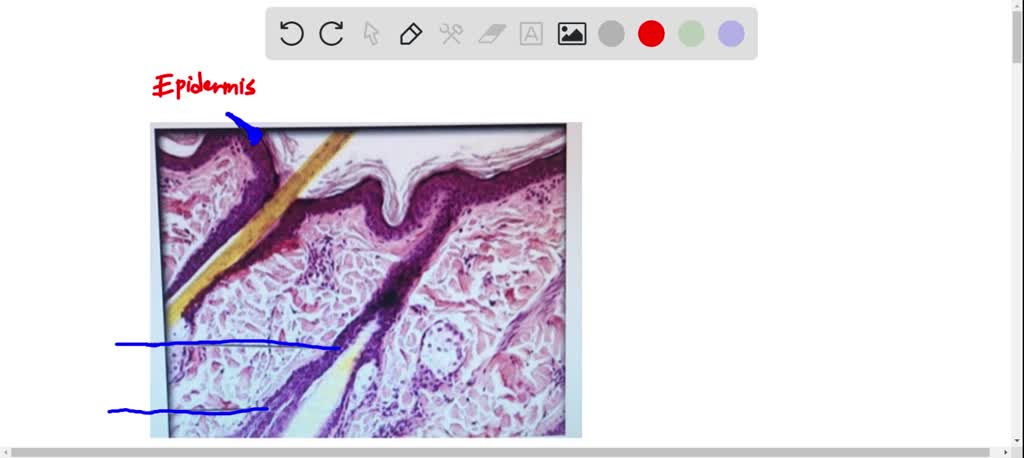
Layers of the Skin | Anatomy and Physiology I - Lumen Learning The skin is composed of two main layers: the epidermis, made of closely packed epithelial cells, and the dermis, made of dense, irregular connective tissue that houses blood vessels, hair follicles, sweat glands, and other structures. Beneath the dermis lies the hypodermis, which is composed mainly of loose connective and fatty tissues. Label the photomicrograph of thin skin. Dermis Duct of sebaceous gland ... Dermis Duct of sebaceous gland Hair Follicle Sebaceous gland Hair Epidermis 1 See answer Advertisement chemisst The skin is a critical organ that covers the entirety of the body's exterior and functions as a barrier that defends the body from viruses and injuries that can be caused by the surrounding environment.
C ezto.mheducation.com/hm.tpx 23. Label the photomicrograph of thin... Label the photomicrograph of thin skin. Hair Sebaceous gland Dermis Hair Follicle Epidermis Duct of sebaceous gland KS Name the structure. Type here to search T... Biology Science Anatomy Answer & Explanation Solved by verified expert All tutors are evaluated by Course Hero as an expert in their subject area. Answered by Lieutenant_jefferyi Answer:

Label the photomicrograph of the sebaceous gland.
anatomy lab, exam 3, lab 9, Spinal Nerves, Integument, and ... - Quizlet Name the highlighted nerve that provides some of the innervation to joints of the hands. median nerve Label the structures of the skin and subcutaneous tissues. epidermis hair hypodermis sebaceous gland dermis deep fascia nerve Match the label to its appropriate spinal cord component. grey matter white matter denticulate ligament dorsal ramus Label the photomicrograph of thin skin. Dermis Duct of sebaceous gland ... Dermis Duct of sebaceous gland Hair Follicle Sebaceous gland Hair Epidermis. We store cookies data for a seamless user experience. ... Label the photomicrograph of thin skin. Dermis Duct of sebaceous gland Hair Follicle Sebaceous gland Hair Epidermis. 1 Approved Answer. KARRI S answered on June 09, 2021. Label the photomicrograph in Figure 7.4. Examine a slide of hairy skin ... Label the structures of the skin in this micrograph by clicking and dragging the labels to the correct location. Activity 4 Differentiating Sebaceous and Sweat Glands Microscopically Using the slide thin skin with hairs and the photomicrographs of cutaneous glands (Figure 7.6) as a guide, identify sebaceous and eccrine sweat glands.
Label the photomicrograph of the sebaceous gland.. Question : Label the photomicrograph of the sebaceous gland. - Chegg Question: Label the photomicrograph of the sebaceous gland. This problem has been solved! You'll get a detailed solution from a subject matter expert that helps you learn core concepts. See Answer Show transcribed image text Expert Answer 100% (31 ratings) Transcribed image text: Label the photomicrograph of the sebaceous gland. Sebaceous Gland Label The Photomicrograph Of Thin Skin - Blogger Sebaceous Gland Label The Photomicrograph Of Thin Skin - Integumentary System Histology Post a Comment And lymph vessels, nerves, and other structures, such as hair follicles and sweat glands. Using the slide thin skin with hairs, and the photomicrographs of cutaneous glands (figure 7.7) as . This problem has been solved! PreLab03a Integument & Prelab03b Integument Histology Label the structures of the skin and subcutaneous tissues. Left side form top: epidermis sebaceous dermis dermis deep fascia nerve Right side from top: hair hypodermis Organize the following layers of epidermis from superficial too deep. stratum corneum stratum lucidum stratum granulosum stratum spinosum stratum basale Figure 7.4 Photomicrograph of the skin and accessory structures - Quizlet Sebaceous Gland Oil glands that surround hair follicles; secrete oils that lubricates skin, hair, and into the neck of the hair follicle. Hair Follicle Surrounds the hair root; formed from epidermal layers that project into the dermis Hair Root extends into the dermis of skin & sometimes the hypodermis
[Solved] . Which structure is highlighted?. Which structure is ... Which structure is highlighted? Dermal papillae Victor Eroschenko Label the photomicrograph of the sebaceous gland. Hair follicle Hair follicle Duct of sebaceous gland Sebaceous gland Sebaceous gland Basal cell Duct of Basal cell sebaceous gland Secretory cell Secretory cell. Identify the structure that is indicated by the leader line and ... Solved Label the photomicrograph of the skin and its | Chegg.com Label the photomicrograph of the skin and its accessory structures. Sebaceous gland Duct of sebaceous gland Epidermis Hair follicle This problem has been solved! You'll get a detailed solution from a subject matter expert that helps you learn core concepts. See Answer Question: Label the photomicrograph of the skin and its accessory structures. Sebaceous Glands: Function, Location & Secretion Sebaceous glands are microscopic glands found in your hair follicles that secrete sebum. Sebum is an oily substance that protects your skin from drying out. Sebaceous glands can clog, so you can keep your glands healthy by following a skin care routine that includes cleansing and moisturizing your skin. Function. Anatomy. Conditions and Disorders. Label the Photomicrograph of the Sebaceous Gland. Solved Label The Photomicrograph Of The Sebaceous Gland Chegg Com Organize the following layers of the epidermis from superficial to deep. The skin consists of two layers. Boys can also blame testosterone from gonadal puberty pubarche. Use Enter Space to view and traverse through the list of languages.
Bio Lab Chapter 6 Quiz Flashcards | Quizlet -hair follicle and sebaceous gland Identify the type of tissue that composes the epidermis of the skin. stratified squamous epithelial tissue Identify the structures of the dermis. dense connective tissue with fibers oriented in many directions dense irregular loose connective tissue characterized by long, thin dark fiber areolar tissue Question: Label the photomicrograph. Myoepithelial cell Lumen ... - Chegg Question: Label the photomicrograph. Myoepithelial cell Lumen Epithelial cell Apocrine sweat gland OMG Den Stree Show transcribed image text Expert Answer 94% (16 ratings) The given micrograph is labelled and is attached below: Justification: Epithelial cell: The epithelial cells in mammary … View the full answer Transcribed image text: Solved Label the photomicrograph of the skin and its | Chegg.com Epidermis Sebaceous gland Hair follicle Duct of sebaceous gland Label the photomicrograph of the skin and its accessory structures. Epidermis Sebaceous gland Hair follicle Duct of sebaceous gland Show transcribed image text Expert Answer 100% (1 rating) Transcribed image text: Label the photomicrograph of the skin and its accessory structures. C ezto.mheducation.com/hm.tpx 23. Label the photomicrograph...ask 8 Helium porosity. Assume that the helium porosity (in percentage) of coal samples taken from a particular seam is normally distributed with a population standard deviation of 0.69. We are interested in constructing a confidence interval for the true average helium porosity (?) in the seam.
Lecture 4 Chapter 5: The Integumentary System - ppt video ...
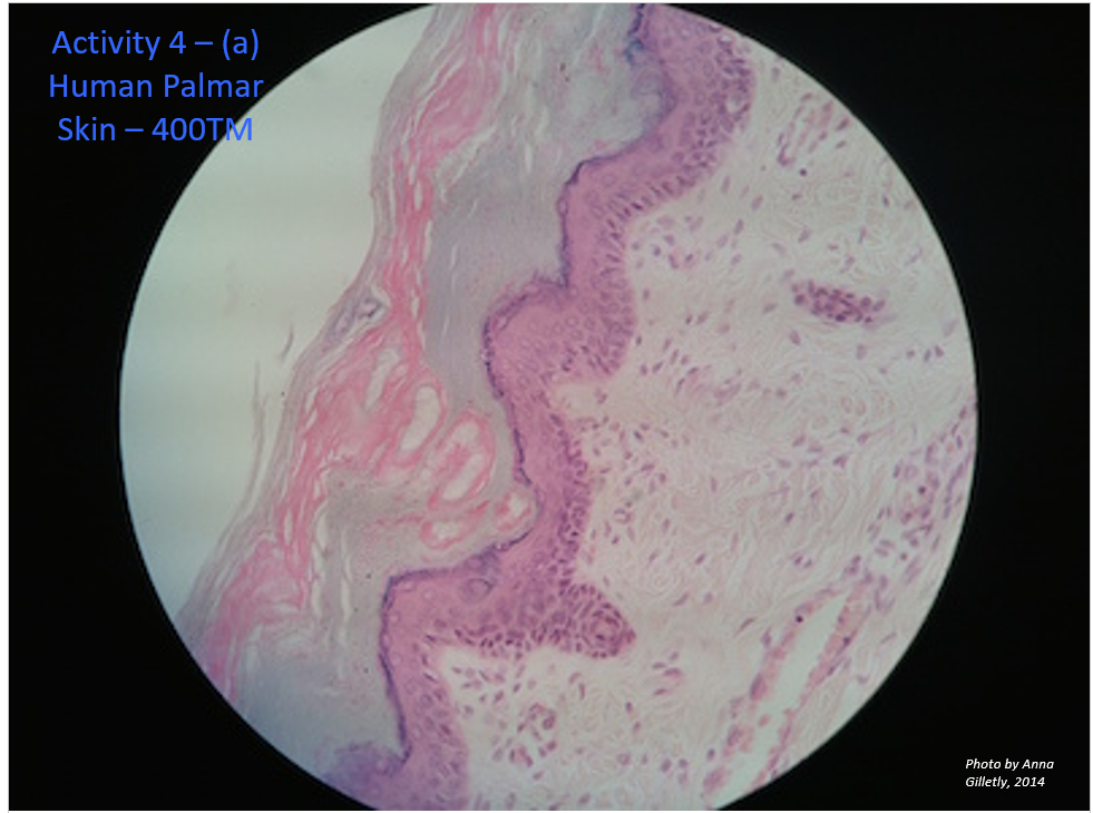
📐Label the photomicrograph of thick skin. Stratum corneum Stratum ... Click here 👆 to get an answer to your question ️ Label the photomicrograph of thick skin. Stratum corneum Stratum basale Stratum granulosum Stratum lucidum Ep…
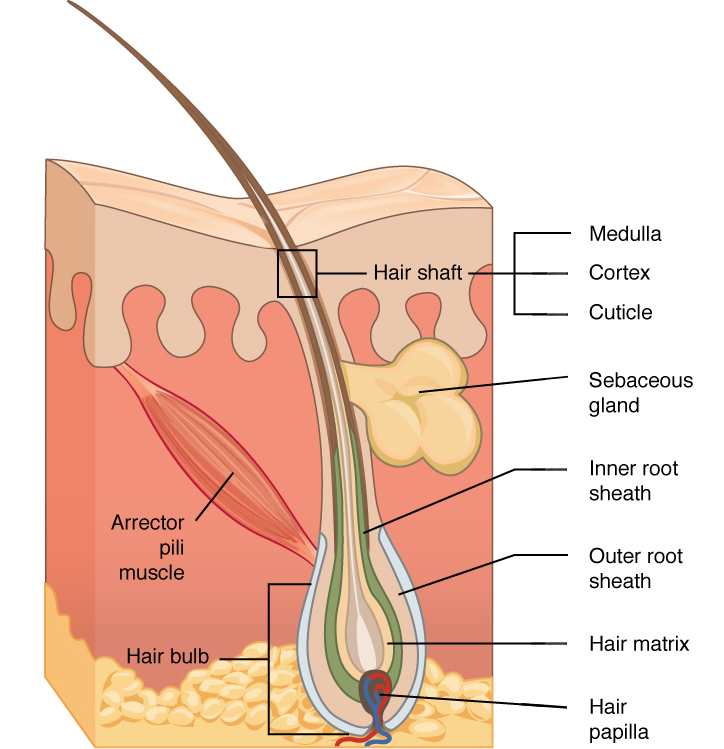
Accessory Structures of the Skin | Anatomy and Physiology I
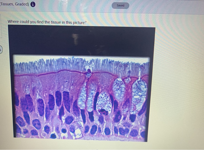
Final Exam A&P 1 Flashcards | Quizlet Label the photomicrograph of thin skin Hair shaft, epidermis, dermal root sheath, sebaceous gland, dermis, hair matrix label the structures of the hair follicle Identify the layers of the epidermis with relation to their location and role in keratinization ... the receptors responsible for olfaction are located in the olfactory epithelium
Solved Label the photomicrograph of the skin and its | Chegg.com
(Solved) - Label The Photomicrograph Of The Skin And Its Accessory ... Label the photomicrograph in Figure 7.4. Examine a slide of hairy skin and identify the structures in Figure 7.4. Hair bulbs hair follicle hair squareroot papilla of hair sebaceous gland. Posted 8 months ago. Q: Art-Labeling Activity: Basic anatomy of the skin Drag the appropriate labels to their respective targets.

Human scalp, light micrograph - Stock Image - C056/7668 ...
Label the photomicrograph in Figure 7.4. Examine a slide of hairy skin ... Label the structures of the skin in this micrograph by clicking and dragging the labels to the correct location. Activity 4 Differentiating Sebaceous and Sweat Glands Microscopically Using the slide thin skin with hairs and the photomicrographs of cutaneous glands (Figure 7.6) as a guide, identify sebaceous and eccrine sweat glands.

Sebaceous Gland Holocrine Gland Whose Cells Foto Stok ...
Label the photomicrograph of thin skin. Dermis Duct of sebaceous gland ... Dermis Duct of sebaceous gland Hair Follicle Sebaceous gland Hair Epidermis. We store cookies data for a seamless user experience. ... Label the photomicrograph of thin skin. Dermis Duct of sebaceous gland Hair Follicle Sebaceous gland Hair Epidermis. 1 Approved Answer. KARRI S answered on June 09, 2021.

BIOL 319 Lab 1 Flashcards | Quizlet
anatomy lab, exam 3, lab 9, Spinal Nerves, Integument, and ... - Quizlet Name the highlighted nerve that provides some of the innervation to joints of the hands. median nerve Label the structures of the skin and subcutaneous tissues. epidermis hair hypodermis sebaceous gland dermis deep fascia nerve Match the label to its appropriate spinal cord component. grey matter white matter denticulate ligament dorsal ramus
Integumentary System Overview
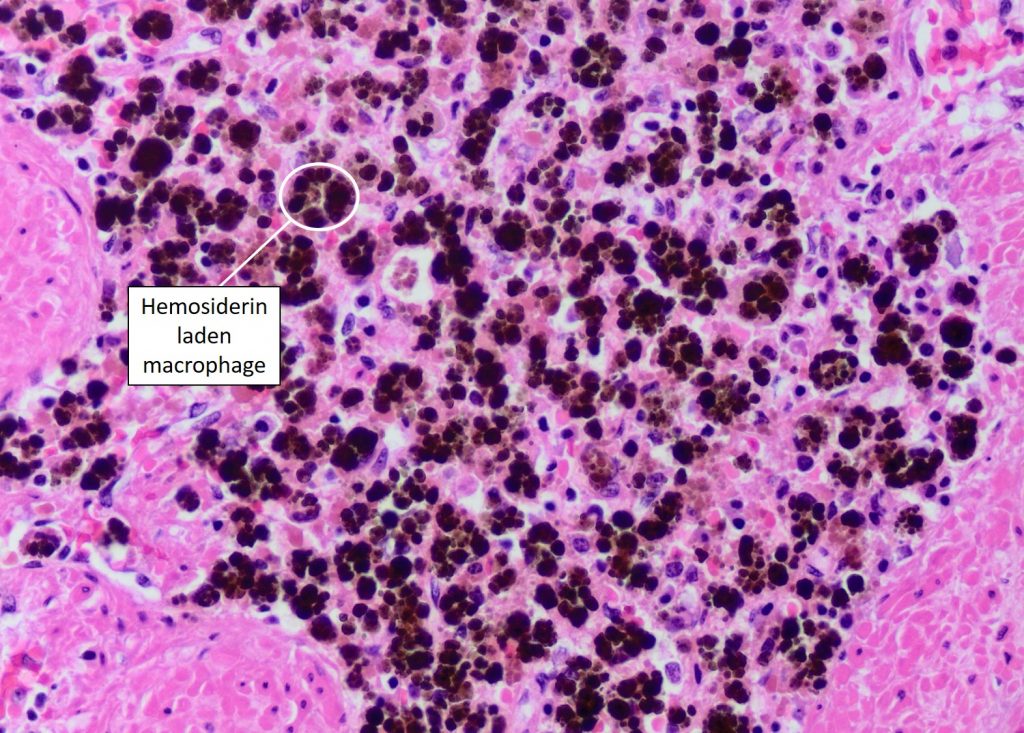
Cell Inclusions – Veterinary Histology
Solved] what are the labels for these? | Course Hero

BIOL 319 Lab 1 Flashcards | Quizlet

Photomicrograph of skin taken from the same animal and ...

Hamza Khan (captainhamzakhan0) - Profile | Pinterest
Answered: What did you notice about the Human… | bartleby

Photomicrograph of group IA showing irregular bone trabeculae ...

Warthin-like mucoepidermoid carcinoma of the parotid gland: a ...

BIO - 168 Final Exam Study Guide Flashcards | Quizlet
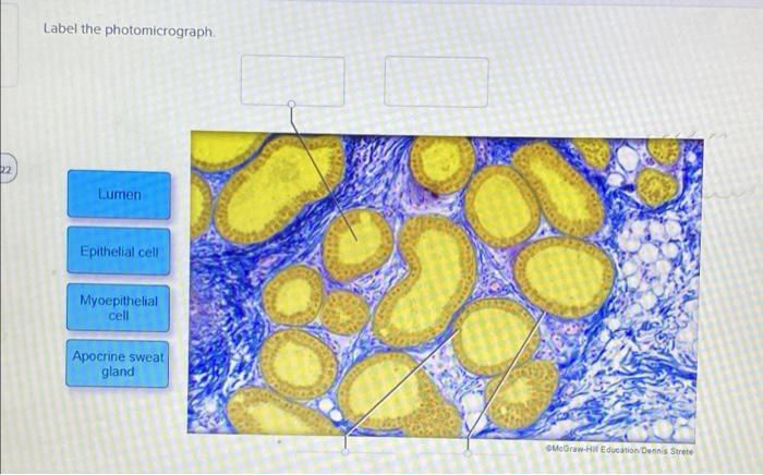
Solved Label the photomicrograph. Lumen Epithelial cell ...

Photomicrograph Of Human Scalp Showing Epidermis Dermis ...
1,726 Immunofluorescent Photomicrograph Photos and Premium ...

Skin and Mammary Gland - ScienceDirect
Integumentary System Overview

View Image
Astragalus membranaceus and Punica granatum alleviate ...

Human scalp, light micrograph - Stock Image - C054/3494 ...

The photomicrograph shows a fibrofolliculoma with the typical ...
Folicle hi-res stock photography and images - Alamy
SISTEM INTEGUMEN [Compatibility Mode]
Solved] Help pls LAB 5 EXERCISE 5-13 In the photomicrograph ...

a) A photomicrograph of the section of thin skin tissue from ...
Infiltrating BCC
Solved Label the photomicrograph of thin skin. | Chegg.com

JaypeeDigital | eBook Reader
Skin and the Integumentary System
Eccrine sweat gland hi-res stock photography and images - Alamy
Establishing human lacrimal gland cultures from biopsy-sized ...
The Cellular Component – Veterinary Histology

Page 2 | Metastasis Images - Free Download on Freepik

Sebaceous hyperplasia of the vulva: a clinicopathological ...
Solved Label the photomicrograph of thin skin. Dermis Duct ...
Stained slide hi-res stock photography and images - Alamy

Label the photomicrograph of the skin and its accessory ...
Label tne photomicrograph Of the Skin and Its accessory structures, Sebaceous gland, Duct ofl, sebaceous gland, Epidermis, Hair follicle
Post a Comment for "42 label the photomicrograph of the sebaceous gland."